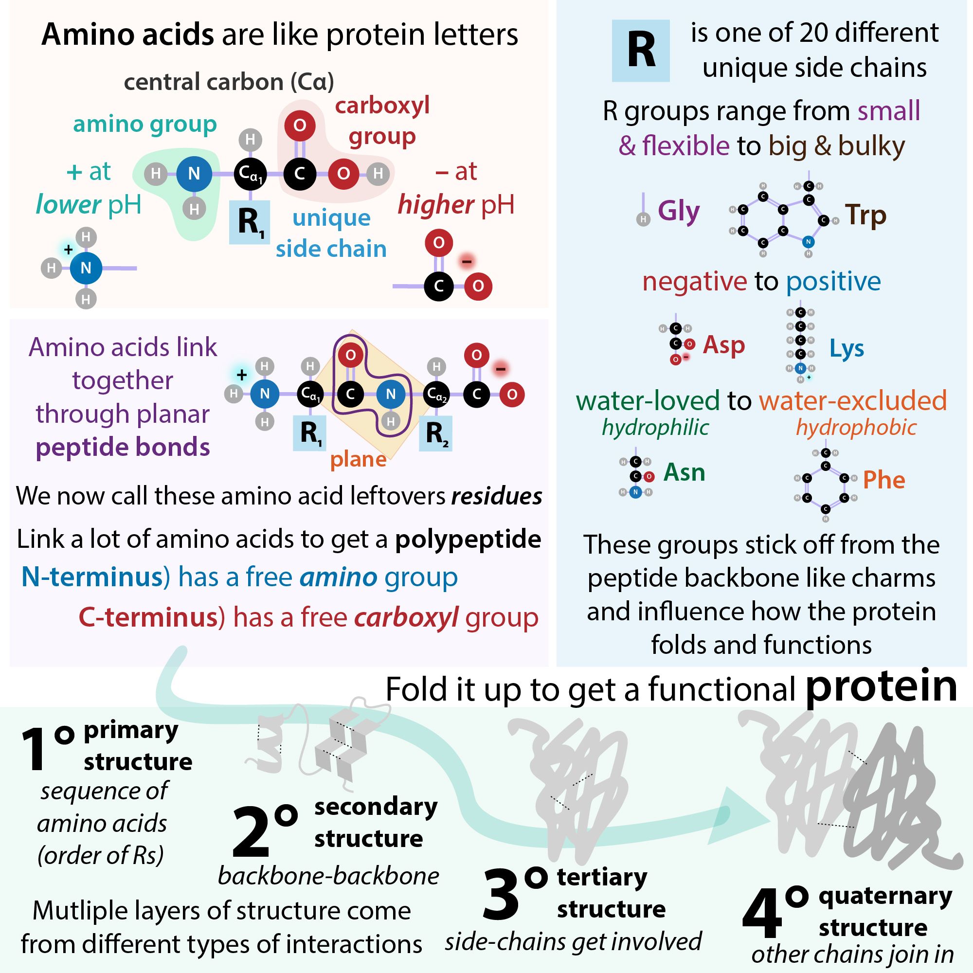
Since Stanley Prusiner discovered prions, so-called “infectious proteins” best known for causing “mad cow disease,” it has become clear that a similar phenomenon as the infectious process of prions occurs in many diseases characterized by the buildup of orderly aggregates of misfolded proteins called amyloids. (Such conditions may collectively be referred to as “amyloidopathies,” a subdivision of “proteinopathies.”) Notably, the two most common neurodegenerative diseases: Parkinson’s Disease (PD) and Alzheimer’s disease (AD) fall into this category, as do others such as Huntington’s disease (HD). So, what’s this phenomenon I’m referring to? As the term “proteinopathy” (protein + disease) suggests, it involves misfolded proteins, but it’s much more orderly (yet “wild” in the “fascinating” sense of the word) than simply things clumping up . . .
You know those psychology experiments where people stand backwards in an elevator and then the person comes in and turns around to fit in? It’s kinda like that. One misfolded protein serves as a seed, a kind of trend-setter, that acts as a template of sorts and causes the others to misfold “to be like it” – though at a biophysical level it’s more that they have to fold that way in order to form favorable interactions. I like to think of it as akin to starting a spooning cuddle chain. Proteins that had previously been healthy, productive cellular denizens suddenly are sleeping on the job. And the big cuddle chain they form gets in the way of other cellular components trying to go about their normal business unless management’s able to intervene.
A bit more scientifically:
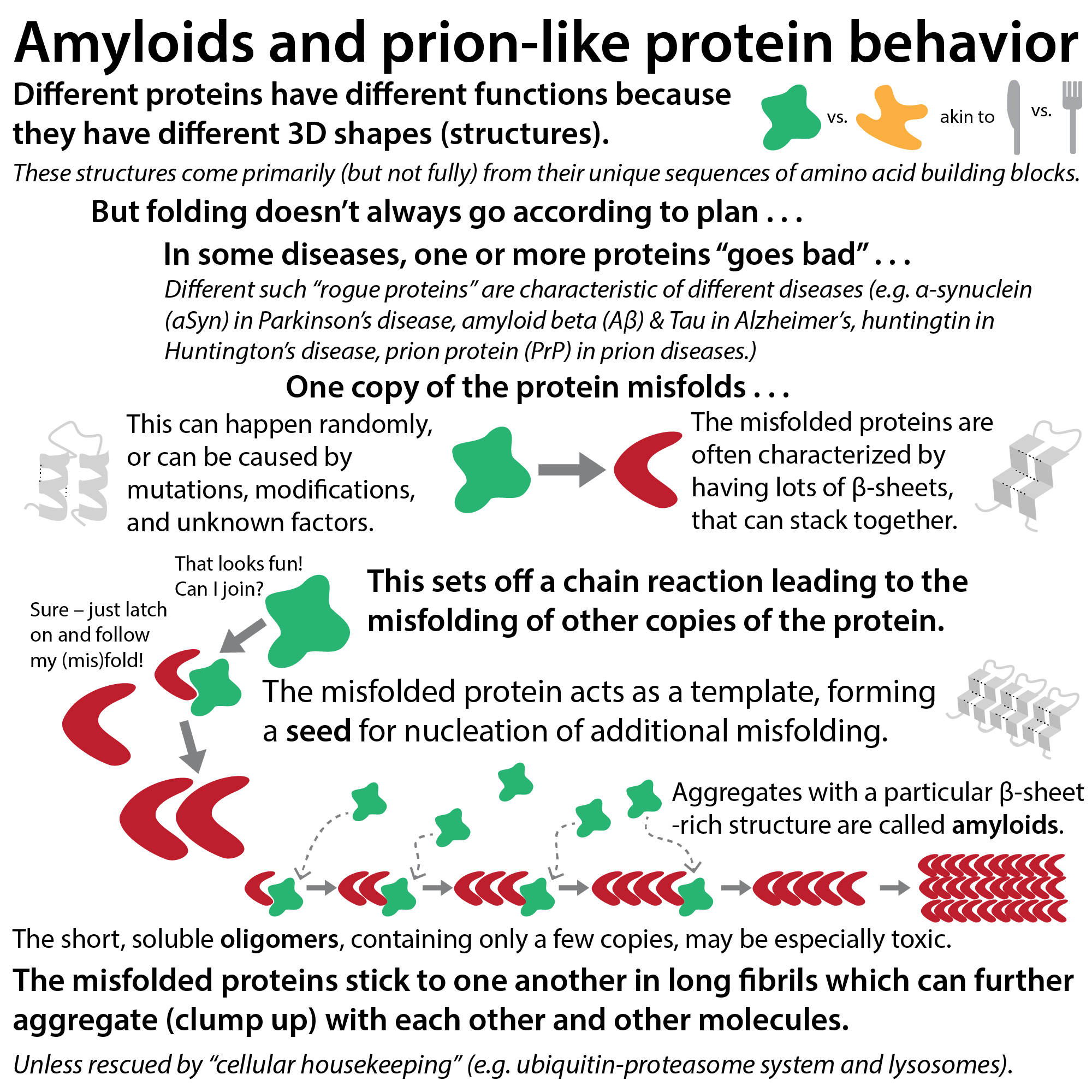
One copy of the protein misfolds → acts as seed, or nucleation point → other copies of the protein similarly misfold, propagating the formation of small groups of misfolded proteins (oligomers, where “oligo” means “few”) and those grow into larger chains called proto-fibrils and then even larger ones called fibrils. Aggregates with a particular structural pattern (rich in stacks of β-sheets*) are called amyloids. These can further aggregate (clump up) with each other and other molecules, unless rescued by “cellular housekeeping” (e.g. ubiquitin-proteasome system and lysosomes). Intriguingly, growing evidence suggests that, at least in some diseases including PD, the soluble oligomers are the most toxic forms and that the larger aggregates could actually be somewhat protective (I like to think of it as sweeping your mess into a pile where you can’t trip over it).
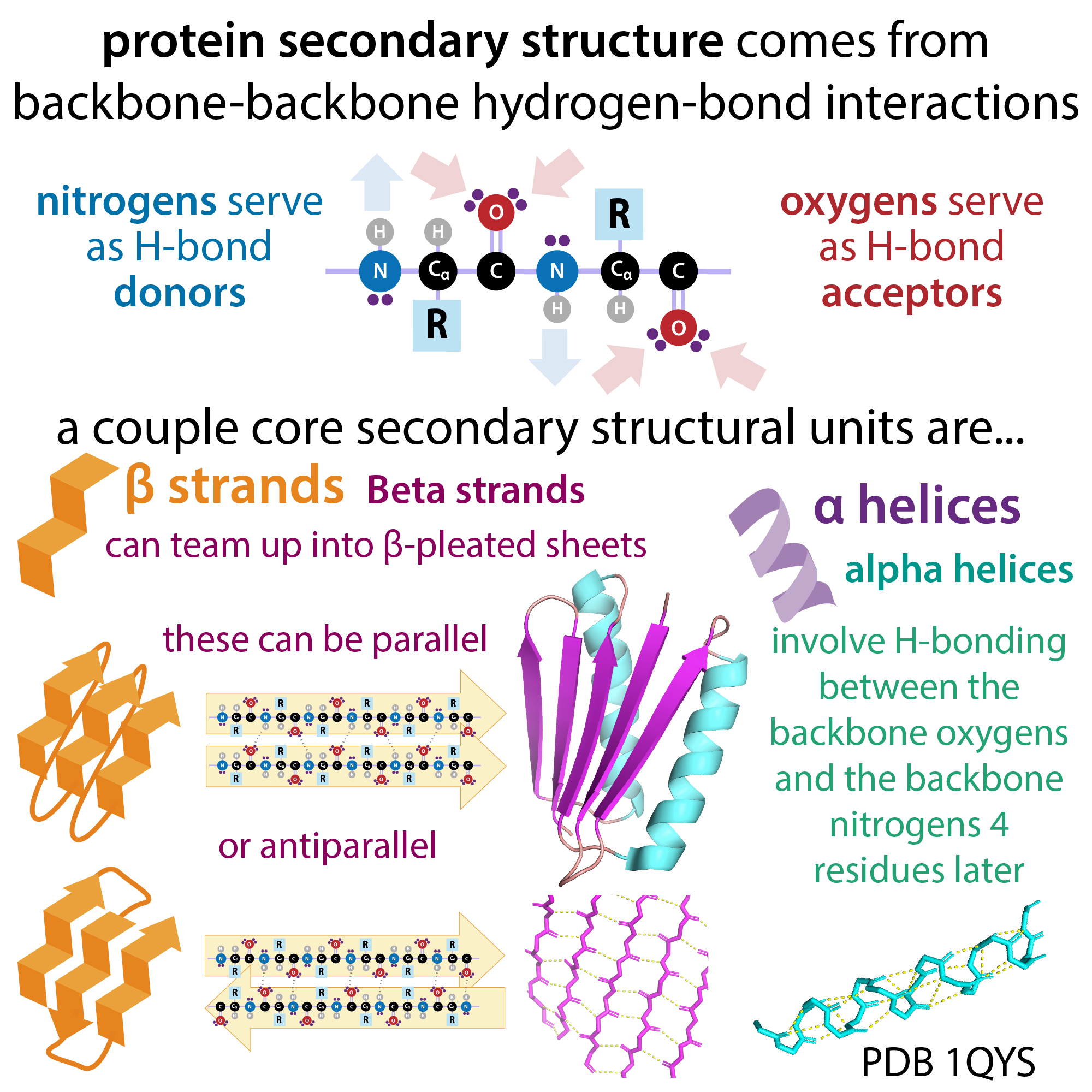
* β-sheets are made up of a common basic protein fold called a β-strand, which is often depicted as a thick arrow. These strands have a zig-zagging backbone (think accordion) that allows them to “snuggle up,” by forming strong hydrogen-bonding interactions with one another. In amyloids the strands are in a so-called “cross-β” orientation, in which the sheets are perpendicular to the axis (think stacks of arrows rather than arrow to arrow).
This propagating protein folding phenomenon was first discovered in an infectious form – that of “prion diseases” such as the ones that cause “scrapies” in sheep, bovine spongiform encephalopathy (“mad cow disease”) in cattle, chronic wasting disease (CWD) in deer, and Creutzfeldt-Jakob disease in humans. The term “prion” (pronounced pre-on, differentiating it from a type of seabird called a “prion” pronounced pr-eye-on), was coined by Dr. Stanley Prusiner in 1982. An amalgamation and slight scrambling of parts of pro(teinaceous) in(fectious particle), he used the term to describe an infectious agent with protein-like physical properties (e.g. light absorbance) that was impervious to disinfection techniques that destroy nucleic acids (DNA and RNA), lipids, and whatever else they threw at it, except intensive protein-destroying methods.
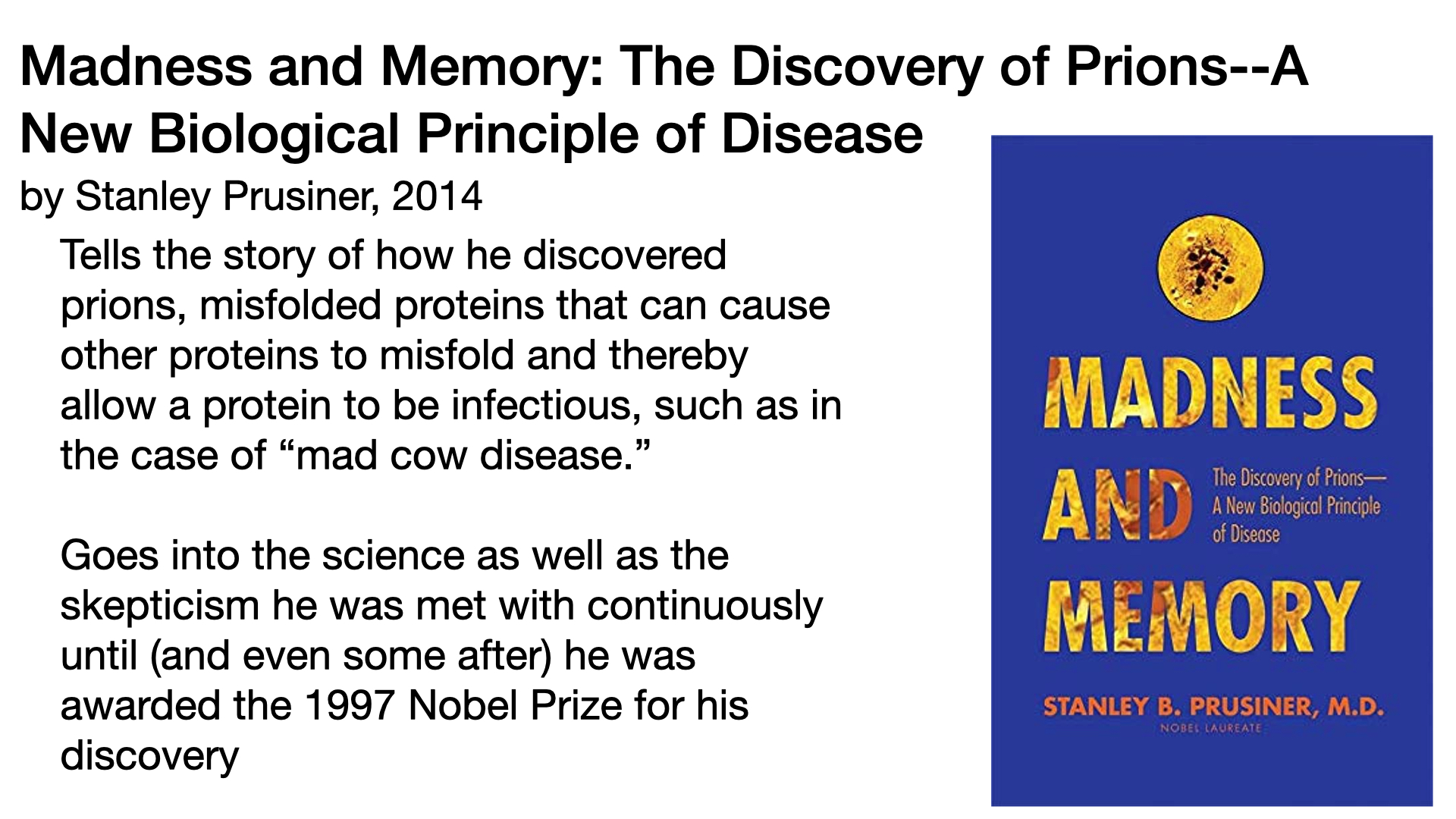
Although most in the field (and most in the world, really) thought his idea was preposterous, Stanley and his team were able to show that the infectious agent they’d identified really was protein! Ultimately, most people came around to it, and he was rewarded the Nobel Prize for Physiology or Medicine in 1997 (I highly recommend Prusiner’s autobiographical telling, “Madness and Memory.”)
Turns out that misfolded copies of distinct proteins in the brains and nervous systems of infected animals could not only cause misfolding of other copies of proteins throughout cells in the infected animals’ bodies, but they could also cause proteins to misfold in the animals that consumed them. Take the “mad cow disease” fiasco of the 90s-early 2000s. Turns out that cattle were being fed infected sheep brains due to relaxed regulatory standards. This led to bovine spongiform encephalopathy (BSE) in the cows and caused variant Creutzfeldt-Jakob disease (vCJD) in some humans who ate contaminated cow tissue.
As their symptoms can take years to manifest, such “transmissible spongiform encephalopathies” are inherently difficult to both track and study in the lab. But scientists including Prusiner were able to develop laboratory models and experimental techniques that shorten the timespan of experiments involving them. This led to a rapid expansion of research on prions and the discovery of “prion-like” behavior at the root of (or at least correlated with) a number of diseases, including several prominent neurodegenerative diseases (diseases that cause gradual destruction of the parts of the nervous system).
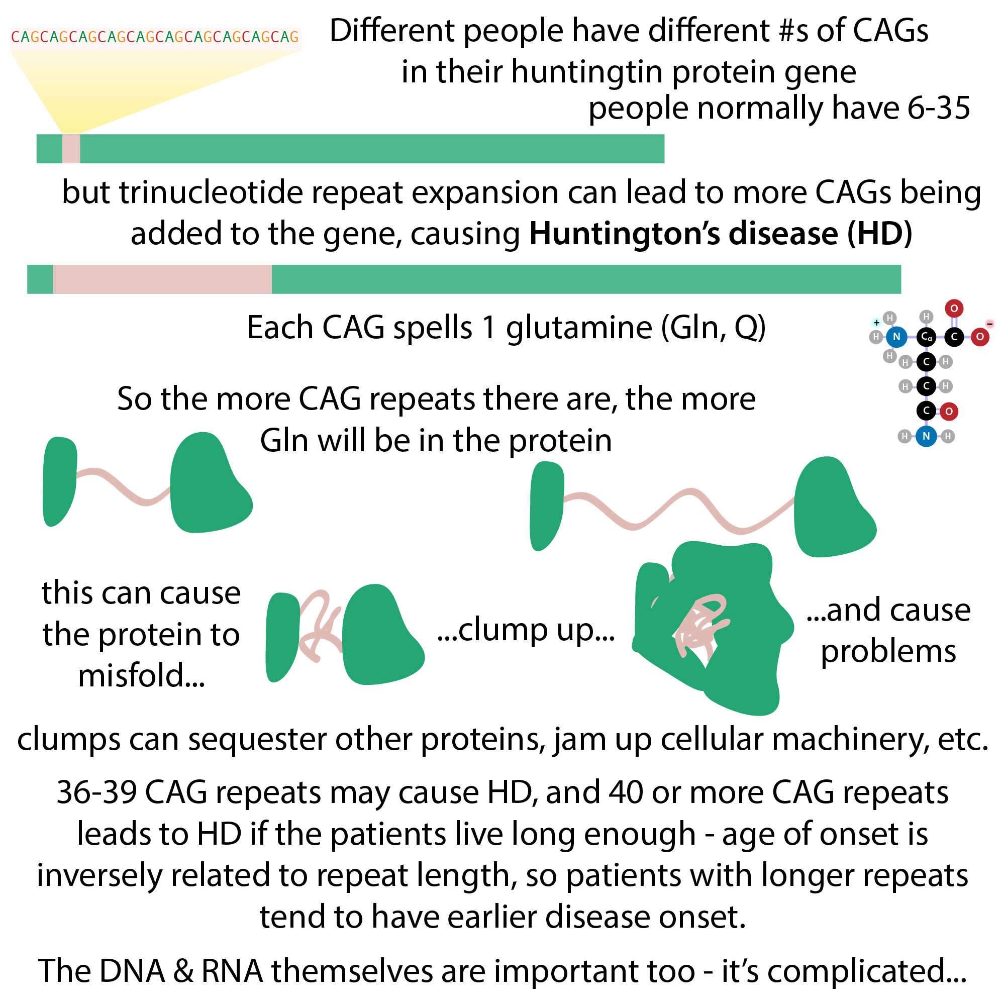
Although their propagation mechanism is the same (or at least very similar), different “rogue proteins” as I like to think of them are characteristic of different such diseases. For example, misfolded α-synuclein (α-syn) is involved in Parkinson’s disease (PD) and other α-synucleinopathies (e.g. Dementia with Lewy bodies (DLB), Multiple System Atrophy (MSA)); amyloid beta (Aβ) and Tau aggregates form in Alzheimer’s disease; huntingtin protein forms amyloids in Huntington’s disease (HD), and the aptly named “prion protein” is at the root of prion diseases.
One thing these proteins have in common is that their “healthy” forms (sometimes denoted with a superscript “c” for cellular) typically have large flexible regions (sometimes referred to as intrinsically-disordered), allowing them to easily “shape-shift” (or, in scientific terms, undergo conformational changes). This allows them to carry out different roles depending on cellular needs (often triggered by binding to a partner protein or lipid or having a chemical modification added). But it also makes them susceptible to misfold when confronted with the protein “peer pressure” of misfolded copies (which are sometimes denoted with a distinguishing superscript, such as “Sc” for “scrapies” in the case of the prion protein, PrP).
For example, with PrP, you get something like:
PrPc + PrPp → PrPp · PrPp
What distinguishes amyloids from other types of misfolded proteins is that although we refer to them as “misfolded,” it’s more like they’re “alternatively-folded.” More akin to a crystalline lattice than to boiled egg whites. Rather than simply wadded up randomly (think of tossing sweaters in your suitcase), they’re folded, just in an odd way (think of folding them inside out or something). And all of them are folded in the “same way” so that they fit together nice and snuggly (making them highly resistant to destruction). These “alternatively-folded” amyloids have a large amount of β-sheet structure, which is particularly amenable to those types of interactions (as compared to, say, the α-helical (spiral staircase like) nature of some protein folds), allowing them to form chains of various lengths. Hence, we can refer to the initial misfolded protein copies as “seeds” that can nucleate the development of soluble oligomers and then fibers and larger insoluble aggregates. A bit like those instant hot packs where you snap a piece of metal and it crystalizes. Although the initial misfolding can be a chance event you wait ages for (though typically it’s more common in disease conditions), once it starts, it grows rapidly.
There are often distinct names given to amyloids of different types and locations. In our α-synucleinopathies example, “Lewy bodies” (LBs) is the name used to refer to amyloids of α-syn (along with phosphorylated tau (p-tau), and amyloid beta protein (Aβ)) in the body of neurons (the classical brain cells that communicate with one another). Similar aggregates, when found in the “arms” (axons and dendrites) of neurons are called Lewy neurites. In contrast to Lewy bodies, which are found in neurons, glial cytoplasmic inclusions (GCIs) sometimes referred to as Papp-Lantos bodies, while also made up of α-Syn aggregates, are found a different type of cell of the nervous system, glial cells, which are often thought of a bit like “helper cells.”
Even the “same” type of aggregates can cause (or at least be associated with) wildly different consequences, depending on their location inside of cells and inside of the body. So much so that differences in localization often differentiate between different diseases. For example, Lewy bodies are characteristic of PD and DLB, whereas GCIs are characteristic of MSA.
MSA involves different cell types than PD and DLB, so it might not be that much of a surprise that although they share some symptoms, they differ greatly in many others. For example, unlike PD and DLB, MSA largely causes problems with coordination (i.e. ataxia) and the autonomic nervous system (blood pressure regulation, etc.). But how about PD and DLB? Both involve Lewy bodies, but PD (although it often does affect cognition, especially at later stages) typically begins with a prominent motor component (causing problems with movement), whereas DLB jumps straight to dementia and other cognitive symptoms, in addition to motor symptoms, and progresses more rapidly. What might explain this? In PD, Lewy bodies (LB) form in the dopaminergic (dopamine-producing) neurons of a part of the brain called the substantial nigra pars compacta (SNpc), whereas in DLB, they also form in a part of the brain called the neocortex.


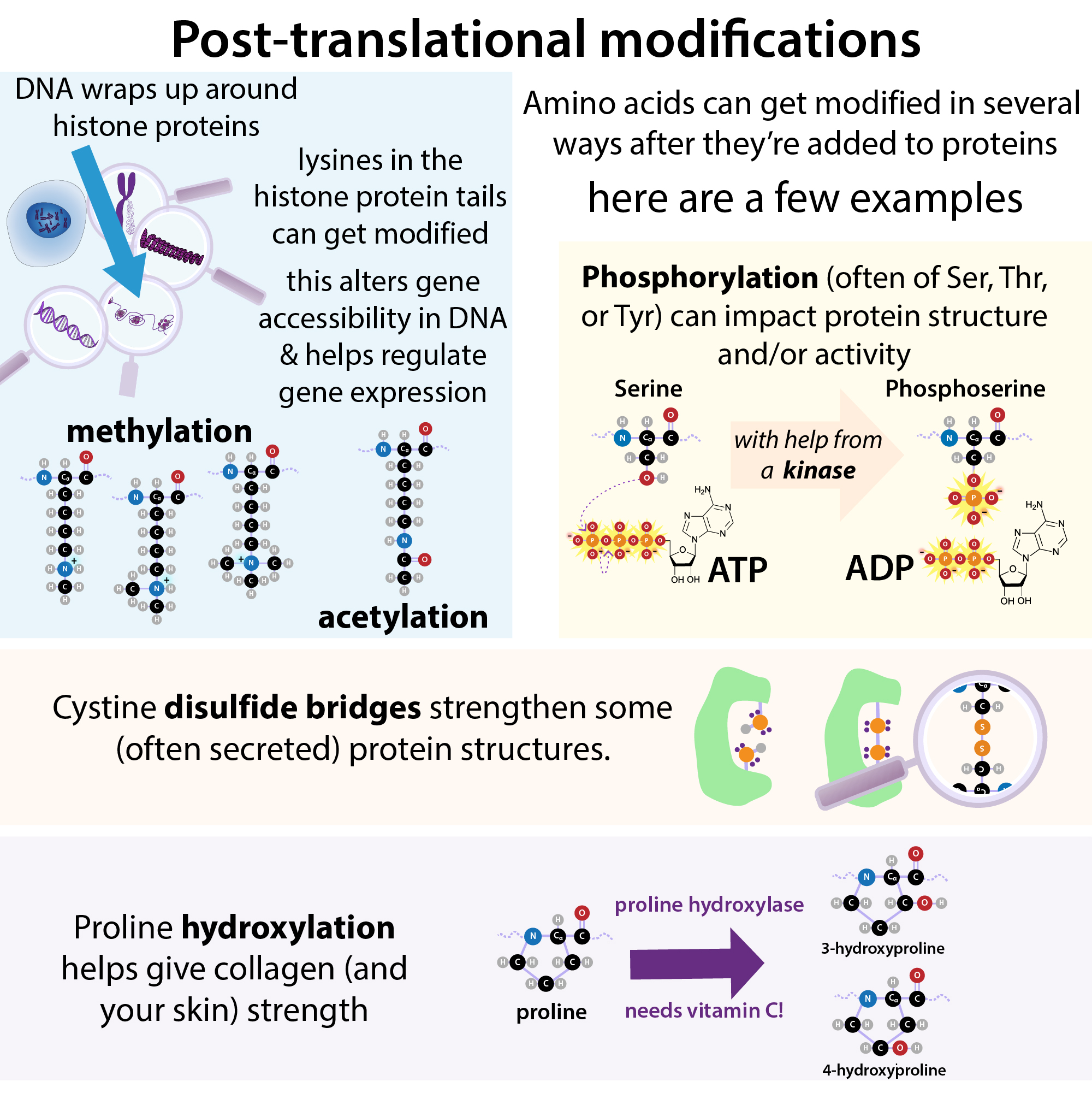
Intriguingly, even for diseases caused by the “same” protein misfolding, evidence is mounting that that “same” misfolded protein actually has slightly different folds (polymorphs), leading to the idea of “strains.” Recall how the healthy proteins usually have large flexible regions that allow them to easily change shape? In addition to protein-protein interactions, and modifications to the protein itself after its amino acids have been pieced together (i.e. post-translational modifications like phosphorylation, which adds a bulky, negatively-charged group to the protein’s surface), the general environment the proteins are in can influence just what shape they’ll change into. This can include things like salt concentration, pH, the presence of other molecules, etc.
This has both complicated the study of these proteins in the lab but also raised the intriguing possibility that differences in the conditions inside of different types of cells, different points in a cell’s life, cells of different people, etc. might influence the development and propagation of different amyloid-related diseases. It could, for instance, help explain why, although there are rare cases of inherited versions of these diseases, involving mutations in the genes for the proteins involved (e.g. the SNCA gene which contains instructions for (encodes) the α-syn protein), the vast majority of cases of PD are “idiopathic” (meaning there’s no clear genetic linkage). It’s tantalizing to think that the existence of different cellular conditions conducive or non-conducive to mis-folding could help account for this.
What’s even more intriguing is that, even under identical laboratory conditions, misfolded proteins from patients with different types of amyloid-related diseases often take slightly different forms (those polymorphs or “strains”) that can be distinguished by different laboratory tests. And some evidence shows that some such “strains” are more toxic, prefer different cell types, etc. This is a hot body of research, and I’ve attached some articles at the end that might be of interest.
Because the large aggregates are such an iconic and glaring aspect of amyloid-related diseases, much therapeutic effort has been aimed at breaking them up, largely to no great success. Why might this be? One theory is that you’re just too late to the game at that point. That might be part of what’s going on, but growing evidence shows that, at least in some diseases, it’s actually the soluble oligomers (those chains of just a few proteins) that cause the most damage.
For example, one mechanism in the case of PD may involve oligomers messing with cellular membranes (e.g. through oligomeric pores). α-syn has a membrane-associating region that allows it to dock on to cellular membranes, and the oligomers can make these membranes leaky.
In some situations, the large aggregates might actually be protective (going back to the idea of sweeping the mess into a corner so you don’t trip on it).
For a lot of these things, it’s not exactly clear how or why in many cases and its disease dependent. But it has huge implications as to what to target with therapies…
In addition to trying to tease apart those and other hows and whys in the lab, scientists are also doing observational studies tracking the progression of the diseases along with corresponding changes in quantifiable biochemical signals, in a search for “biomarkers” that can reliably report on disease state. Scientists hope this knowledge will help them determine how best to catch the diseases early, track them, and intervene. Until then, treatment is restricted mainly to alleviating symptoms, rather than stopping progression.
More on some of the techniques used to study amyloids (especially seed amplification assays) in a future post. For now, hope this helped!
Update: here’s that other post: PCR for Problematic Proteins? Seed Amplification Assays (SAAs)(e.g. PMCA & RT-QuIC) for measuring amyloid precursors and formation
Note: this post is the result of me wanting to understand my dad’s Parkinson’s Disease, help explain it to him, and see what potential future treatments await (hence me focusing most on PD).
Bibliography
- Madness and Memory: The Discovery of Prions–A New Biological Principle of Disease, by Stanley Prusiner, 2014
- Tells the story of how he discovered prions, misfolded proteins that can cause other proteins to misfold and thereby allow a protein to be infectious, such as in the case of “mad cow disease.” Goes into the science as well as the skepticism he was met with continuously until (and even some after) he was awarded the Nobel Prize for his discovery
- Review articles on α-synucleinopathies
- Molecular mechanisms:
- α-syn focused:
- Lashuel, H. A.; Overk, C. R.; Oueslati, A.; Masliah, E. The Many Faces of α-Synuclein: From Structure and Toxicity to Therapeutic Target. Nat Rev Neurosci 2013, 14 (1), 38–48. https://doi.org/10.1038/nrn3406.
- Has some nice graphical summaries in addition to information
- Calabresi, P.; Mechelli, A.; Natale, G.; Volpicelli-Daley, L.; Di Lazzaro, G.; Ghiglieri, V. Alpha-Synuclein in Parkinson’s Disease and Other Synucleinopathies: From Overt Neurodegeneration Back to Early Synaptic Dysfunction. Cell Death Dis 2023, 14 (3), 176. https://doi.org/10.1038/s41419-023-05672-9.
- Lashuel, H. A.; Overk, C. R.; Oueslati, A.; Masliah, E. The Many Faces of α-Synuclein: From Structure and Toxicity to Therapeutic Target. Nat Rev Neurosci 2013, 14 (1), 38–48. https://doi.org/10.1038/nrn3406.
- Broader-scoped:
- Maiti, P; Manna, J.; Dunbar, G. L. Current Understanding of the Molecular Mechanisms in Parkinson’s Disease: Targets for Potential Treatments. Translational Neurodegeneration 2017, 6 (1), 28. https://doi.org/10.1186/s40035-017-0099-z
- α-syn focused:
- Molecular mechanisms:
- Treatment strategies for PD:
- Stocchi, F.; Bravi, D.; Emmi, A.; Antonini, A. Parkinson Disease Therapy: Current Strategies and Future Research Priorities. Nat Rev Neurol 2024, 20 (12), 695–707. https://doi.org/10.1038/s41582-024-01034-x.
- α-synuclein structure:
- Oliveira, L. M. A.; Gasser, T.; Edwards, R.; Zweckstetter, M.; Melki, R.; Stefanis, L.; Lashuel, H. A.; Sulzer, D.; Vekrellis, K.; Halliday, G. M.; Tomlinson, J. J.; Schlossmacher, M.; Jensen, P. H.; Schulze-Hentrich, J.; Riess, O.; Hirst, W. D.; El-Agnaf, O.; Mollenhauer, B.; Lansbury, P.; Outeiro, T. F. Alpha-Synuclein Research: Defining Strategic Moves in the Battle against Parkinson’s Disease. npj Parkinsons Dis. 2021, 7 (1), 65. https://doi.org/10.1038/s41531-021-00203-9.
- Also goes into aspects of clinical trial development, etc. and outlines challenges & opportunities in the field
- Oliveira, L. M. A.; Gasser, T.; Edwards, R.; Zweckstetter, M.; Melki, R.; Stefanis, L.; Lashuel, H. A.; Sulzer, D.; Vekrellis, K.; Halliday, G. M.; Tomlinson, J. J.; Schlossmacher, M.; Jensen, P. H.; Schulze-Hentrich, J.; Riess, O.; Hirst, W. D.; El-Agnaf, O.; Mollenhauer, B.; Lansbury, P.; Outeiro, T. F. Alpha-Synuclein Research: Defining Strategic Moves in the Battle against Parkinson’s Disease. npj Parkinsons Dis. 2021, 7 (1), 65. https://doi.org/10.1038/s41531-021-00203-9.
- Toxicity of oligomers vs fibrils:
- Review articles:
- Wolfe, K. J.; Cyr, D. M. Amyloid in Neurodegenerative Diseases: Friend or Foe? Semin Cell Dev Biol 2011, 22 (5), 476–481. https://doi.org/10.1016/j.semcdb.2011.03.011.
- Mainly discusses AD & HD
- Bigi, A.; Cascella, R.; Cecchi, C. α-Synuclein Oligomers and Fibrils: Partners in Crime in Synucleinopathies. Neural Regen Res 2023, 18 (11), 2332–2342. https://doi.org/10.4103/1673-5374.371345.
- Wolfe, K. J.; Cyr, D. M. Amyloid in Neurodegenerative Diseases: Friend or Foe? Semin Cell Dev Biol 2011, 22 (5), 476–481. https://doi.org/10.1016/j.semcdb.2011.03.011.
- Key research papers for PD:
- Conway, K. A.; Lee, S.-J.; Rochet, J.-C.; Ding, T. T.; Williamson, R. E.; Lansbury, P. T. Acceleration of Oligomerization, Not Fibrillization, Is a Shared Property of Both α-Synuclein Mutations Linked to Early-Onset Parkinson’s Disease: Implications for Pathogenesis and Therapy. Proceedings of the National Academy of Sciences 2000, 97 (2), 571–576. https://doi.org/10.1073/pnas.97.2.571.
- Winner, B.; Jappelli, R.; Maji, S. K.; Desplats, P. A.; Boyer, L.; Aigner, S.; Hetzer, C.; Loher, T.; Vilar, M.; Campioni, S.; Tzitzilonis, C.; Soragni, A.; Jessberger, S.; Mira, H.; Consiglio, A.; Pham, E.; Masliah, E.; Gage, F. H.; Riek, R. In Vivo Demonstration That α-Synuclein Oligomers Are Toxic. Proceedings of the National Academy of Sciences 2011, 108 (10), 4194–4199. https://doi.org/10.1073/pnas.1100976108.
- Review articles:
More about:
- Protein structure: https://bit.ly/proteinstructure
- Huntington’s disease: http://bit.ly/glutaminehd
- Intrinsically disordered regions: https://bit.ly/proteinflexibility
- All sorts of things: #365DaysOfScience All (with topics listed) 👉 http://bit.ly/2OllAB0 or search blog: https://thebumblingbiochemist.com