A number of diseases, including the common neurodegenerative diseases Parkinson’s Disease (PD) and Alzheimer’s Disease (AD) share a common cellular signature: aggregates (clumps) of orderly-misfolded proteins with an amyloid structure (characterized by sturdy stacks of proteins with an accordion-like fold). Because these form in the brain, they typically aren’t detected until autopsy, which doesn’t do patients any good. Instead, if you want to help patients, you need to be able to detect their formation much earlier, while there’s still time to intervene. Now, therefore, scientists are working to establish tests collectively referred to as Seed Amplification Assays (SAAs) (including Protein misfolding cyclic amplification (PMCA) and real-time quaking-induced conversion (RT-QuIC)) to detect the misfolded protein precursors in cerebral spinal fluid (CSF) from lumbar puncture (spinal tap) fluids. And potentially even from less invasive tissue samples such as skin biopsies.
Although there’s not much that can be done to slow disease progression yet, the hope is that by finding reliable biomarkers (measurable signals of an underlying disease that are specific for the disease state) such as misfolded proteins, scientists may be able to better predict, diagnose, track, and understand the disease at the biochemical levels. And use this knowledge to develop effective treatment strategies to detect the disease before it ramps up and stop its pathology in its tracks before it can wreak havoc. (And objectively measure whether the treatments are working.)
In addition to being able to measure the misfolded protein at the heart of amyloids to serve as a biomarker, tests (often referred to as “assays”) that can both quantify (determine amounts present) and chemically characterize the misfolded proteins are useful for scientists trying to figure out how and why the proteins misfold, and whether candidate drugs may be able to stop their misfolding and amyloid production. Furthermore, these tests might be able to tease apart subtle differences in the misfolding of the same protein involved in different diseases (e.g. PD and Dementia with Lewy Bodies (DLB)) to figure out: 1) which disease a patient has and 2) why the diseases progress with different symptoms and at different rates.
Therefore, I want to tell you about how these tests work. But, in order to do that, I need to step back and tell you a bit more about amyloids. For much more about them, please see my prior post (which, as like this current post, also has a comprehensive bibliography at the end for those wanting to learn even more). https://thebumblingbiochemist.com/365-days-of-science/prions/
Quick side note: my dad has PD, and this post was inspired by me wanting to learn about this test I read about involved in both clinical trials and more fundamental biomedical research, so I’m going to focus mainly on PD.
Although the proteins involved are different in different diseases (e.g. amyloid beta (aβ) and tau in AD and alpha-synuclein (α-syn) in PD), the way amyloids form is remarkably similar. It involves a “prion-like” chain reaction whereby one copy of the misfolded (or, as I like to think of it “alternatively-folded”) protein serves as a template that shows the other copies how to misfold. Rather than simply show them, however, that original misfolded protein offers a physical base on which another copy can latch on and misfold in the same manner (doing so because it allows the copies to form lots of favorable interactions (I like to think of them snuggling)). In this way, the original misfolded protein serves as a “seed” to nucleate the subsequent formation of short chains of copies of the misfolded proteins (referred to as oligomers, where “oligo” means few or several), then long chains (fibrils) that can stick to one another, and to other molecules inside of the cell (proteins, membranes, etc.)
So, where does that initial misfolded protein come from? It could happen randomly/spontaneously, but that’s a very infrequent event in a wild-type (unmutated) protein in the absence of any other modulating factors. But, in patients with various neurological diseases like PD and AD, these misfolded proteins are more common, and their misfolding might be provoked by post-translational modifications (addition of chemical groups such as bulky, negatively-charged phosphate groups to an already-synthesized protein), binding to other cellular molecules, or conducive changes in their surrounding environment.
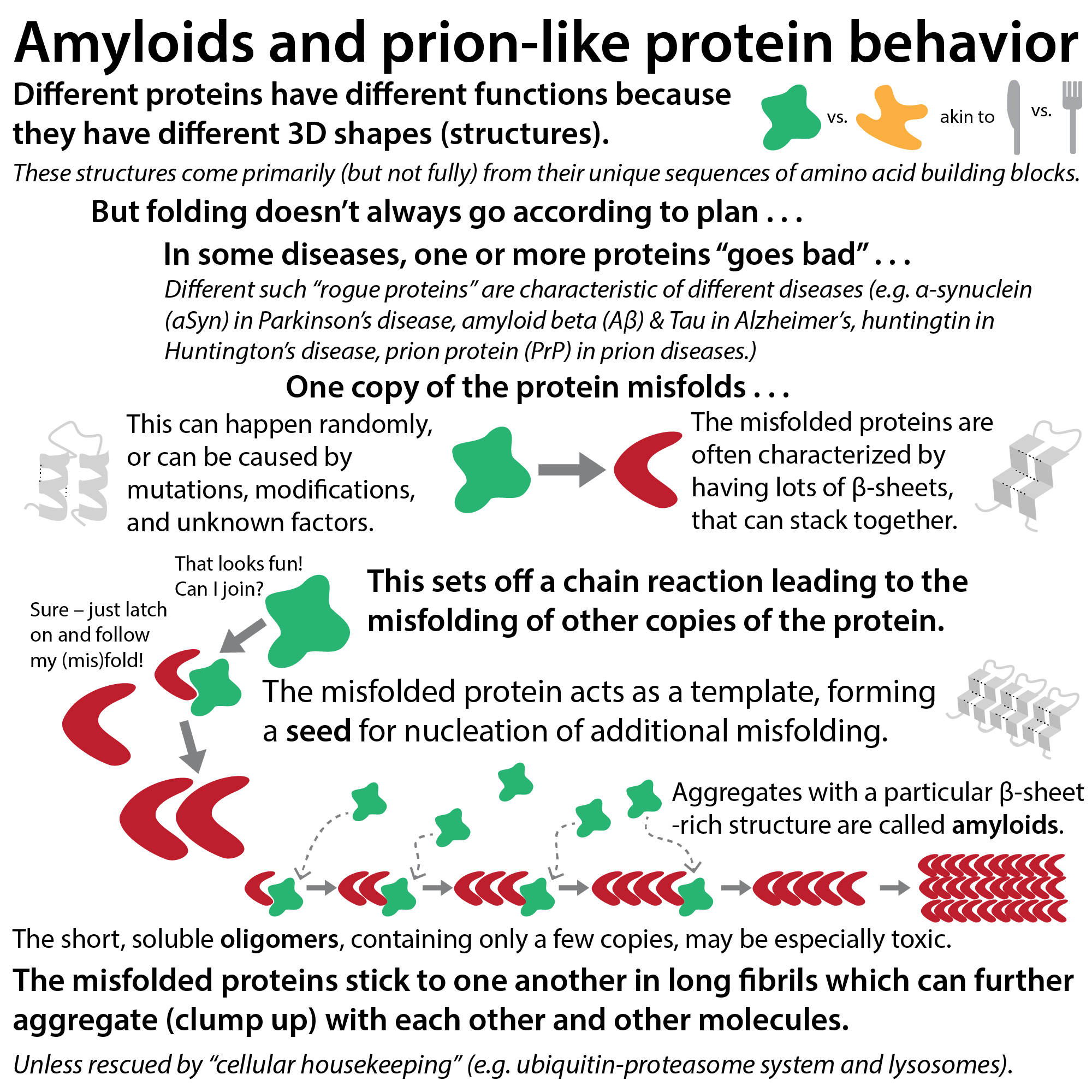
There’s actually still a lot that’s unknown about the factors that trigger seed formation in the cells of afflicted patients (except in more clear-cut, but rare, cases in which patients have a mutation in the gene encoding (containing the instructions for) the protein in question). What is clear, however, is that once a seed forms, fibrils can form with remarkable speed. (Though, remarkably, the actual speed depends on subtle differences in the 3D shape (conformation) of the original misfolded protein seed, leading to the concept of disease-specific “strains” or “polymorphs”). Answering these questions is challenging for a number of reasons, but tests such as SAAs are beginning to provide clues (in addition to laying groundwork for use in diagnostics and tracking of disease progression and/or treatment efficacy).
To discuss these tests, I need to toss around some scientific techno-babble–but don’t let it scare you off; the concept is fairly straightforward! So, let’s start with the concept.
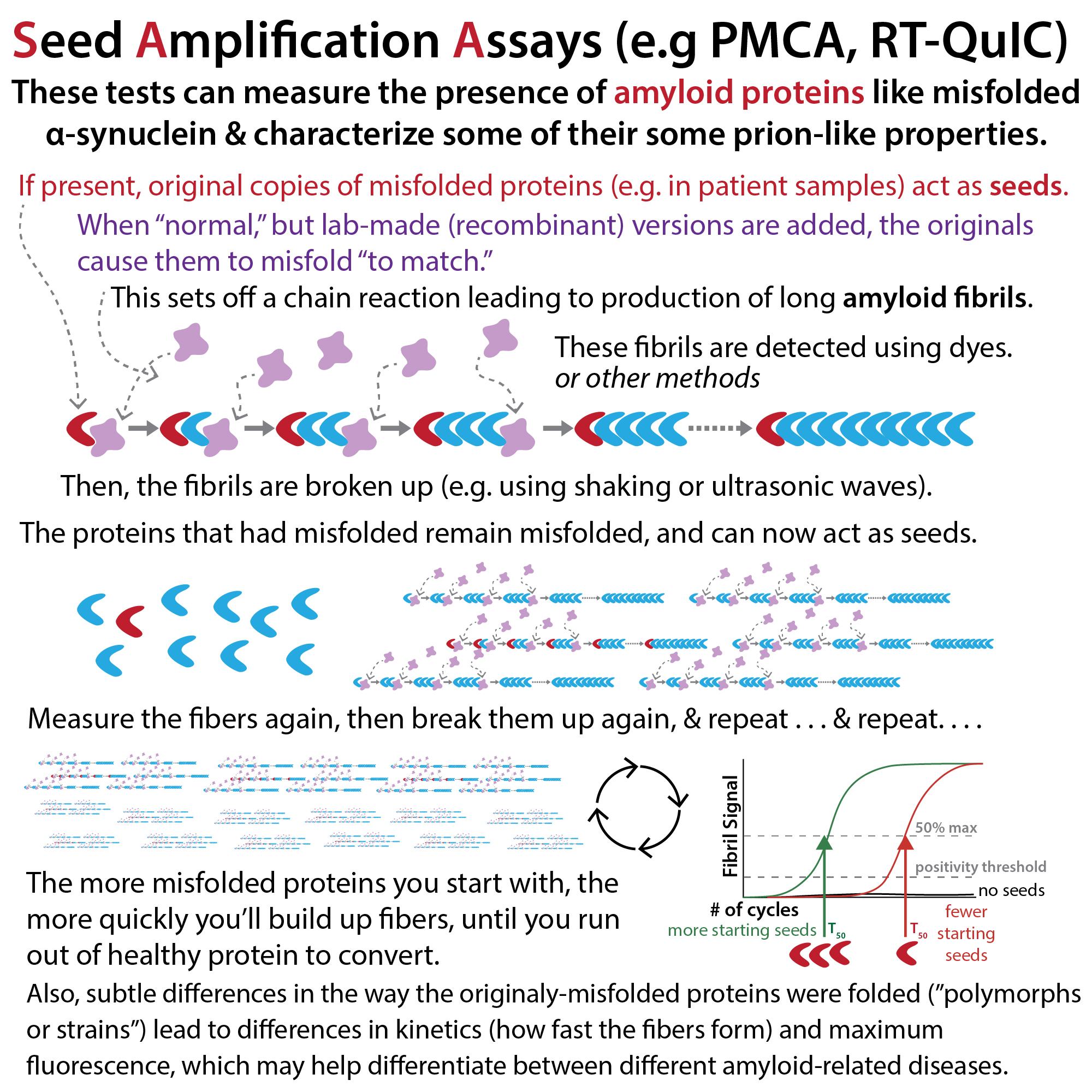
Recall our scenario: misfolded protein “seed” + healthy proteins → soluble oligomer + healthy proteins → fibril of misfolded proteins
In a straight-chain fiber, there’s only one end to grow from (i.e. a single nucleation point). But, if you break up the fibrils, you get back to oligos which can act as nucleation points again. Now, however, you have a lot more of them than before. So, you can amplify the fibril production.
The more misfolded protein copies you had to begin with, the faster you’ll amplify. The fibrils can be detected by methods like dyes that bind them (typically thioflavin-T), and therefore this “break and build” cycling can be used to measure the amount of initial misfolded protein as well as the kinetics (speed) at which the fibrils from the same amount of different misfolded proteins form.
This is the basis for “seed amplification assays” such as Protein Misfolding Cyclic Amplification (PMCA) and Real-Time Quaking-Induced Conversion (RT-QuIC). These tests are really similar (and give consistent results, at least for PD), but they differ in the way the fibers are broken up: PMCA (at least the original version) uses ultrasonic waves in a process called sonication, whereas RT-QuIC literally shakes them). Though I’ve also seen a number of “PMCA” protocols that use shaking, not sonication, so it seems the terminology is a bit murky. Kuang et al. has some tables that compare tons of them.
Although the details vary, scientists typically start by adding lots of copies of a recombinant (lab-made) version of the protein of interest to a test sample (which may be from patient body fluids or another laboratory-made sample). They let it incubate (basically hang out) for a while, in order to enable fibril production. If “proteopathic” copies of the proteins (seeds) were present in the sample, fibrils can form (by recruiting the recombinant protein to the dark side – er, I mean by converting the healthy protein to misfolded protein). But, if pre-formed seeds weren’t present, fibers won’t form (unless a healthy version spontaneously converts which is a very improbable occurrence given energetic barriers). Furthermore, the more seeds that were present originally, the more fibers can form. And the more rapidly the conversions take place, the bigger the fibers can grow. Since there’s only one end per seed to grow from, however, the signal is low.
To get a robust signal, you’ll need a lot more fibrils to form. So, you need a lot more seeds. Thankfully, you can now obtain them by breaking up the fibrils. The more and/or longer the fibrils are when you break them up, the more seeds you’ll get. Give them time to incubate again, measure, break up, incubate, measure, break up, incubate . . . Keep on cycling and eventually the signal will plateau at the maximum fluorescence when all the recombinant protein has been used up. The more and/or or more kinetically active the original sample, the faster your signal will rise and reach that dead end. And, the less time you will need to reach the half maximum fluorescence (T50). The actual maximum fluorescence value will depend in part on how the dye interacts with the aggregates, which depends on the structure of the aggregates (more below) and therefore offers further insight into the particular type of aggregate.
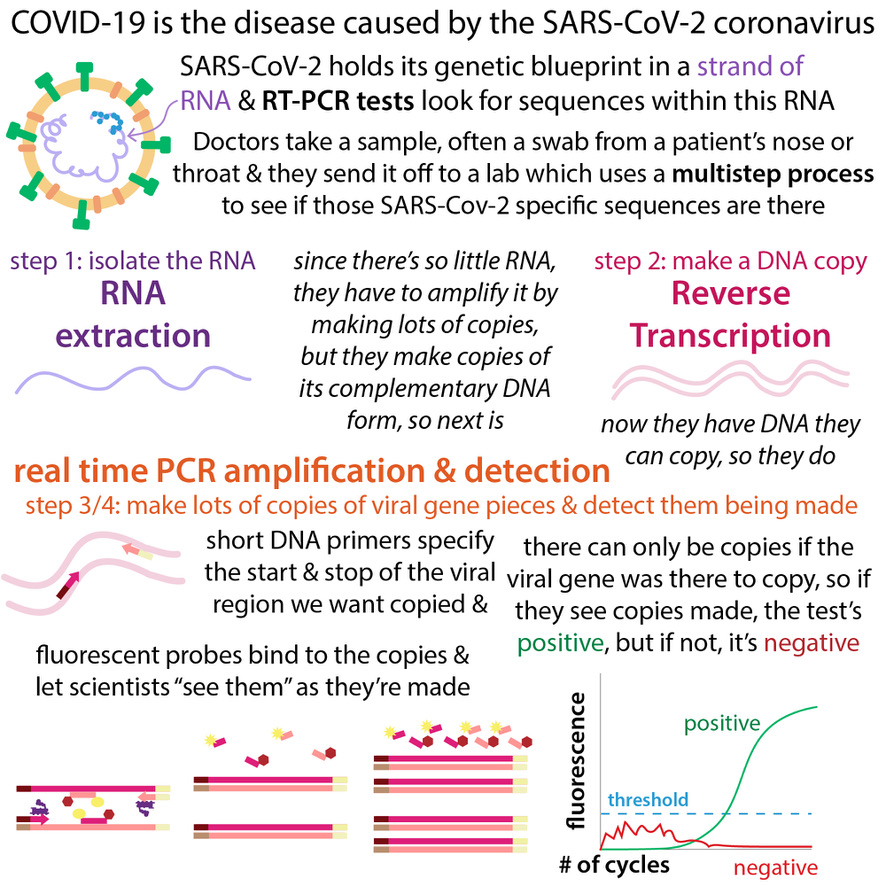
If you’ve heard of qPCR (quantitative Polymerase Chain Reaction), sometimes referred to as RT-PCR (Real-Time PCR), it’s a bit like that. But, in qPCR you’re making copies of DNA and counting the copies (to see how many copies of DNA you started with) rather than making fibrils of proteins (to see how many misfolded proteins you started with). In both cases, however, you carry out the assay in cycles, measuring after each cycle, and your signal grows faster if you start with more. Speaking of more, more on qPCR here: http://bit.ly/rtrtqpcrprimer
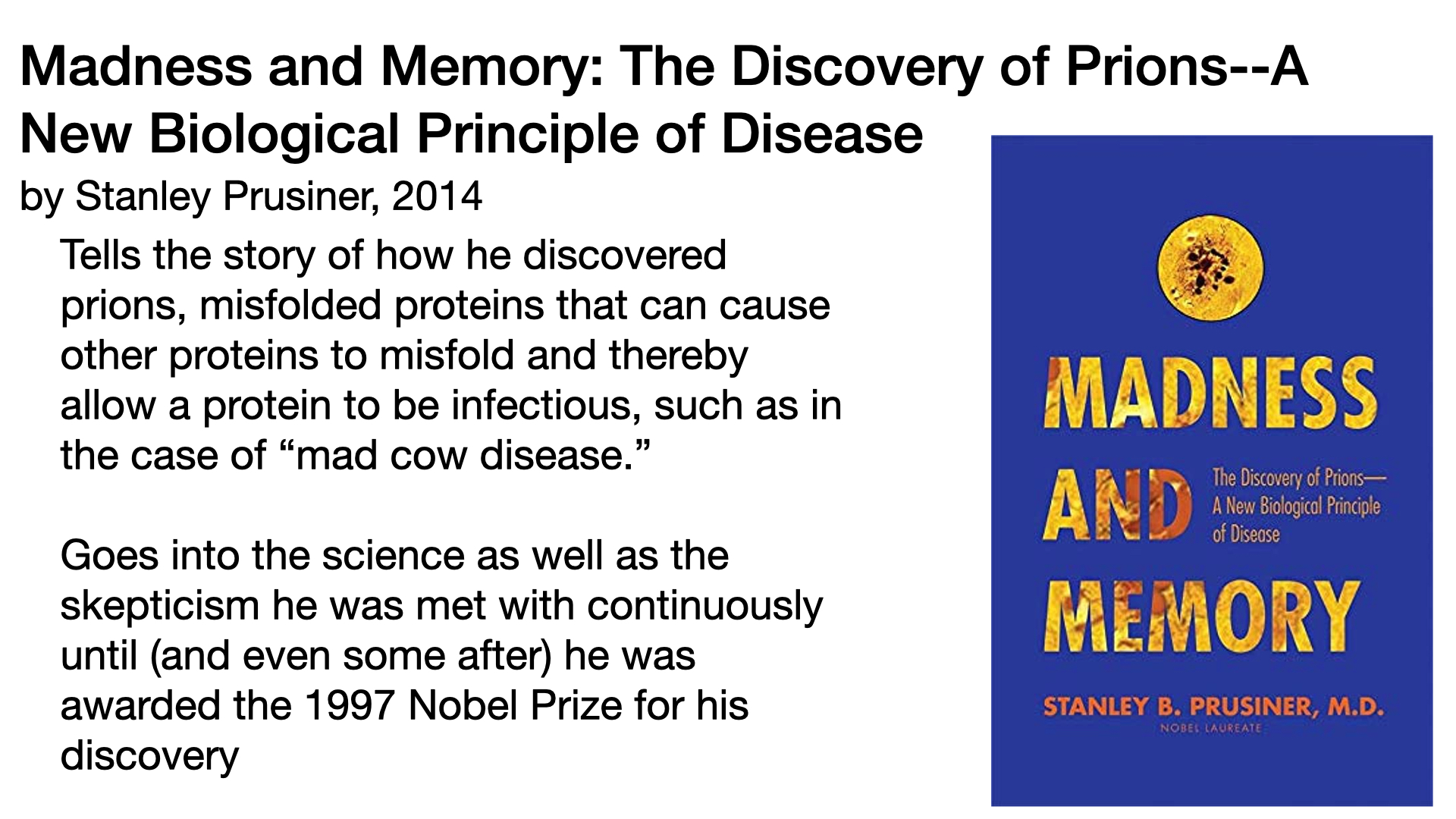
These tests were originally designed for research on prions (pronounced pre-ons), which are infectious proteins that cause (among other diseases) “scrapie” in sheep, bovine spongiform encephalopathy (“mad cow disease”) in cattle, chronic wasting disease (CWD) in deer, and Creutzfeldt-Jakob disease in humans. Those involve different proteins than PD, AD, and other amyloid-involving neurodegenerative diseases, but the amyloid production process is the same, and therefore the tests can be used on them all.
The original versions were low-throughput (you couldn’t test as many things at once) because, instead of dyes they used protease digestion and subsequent gel electrophoresis. Basically, proteases are a type of protein enzyme that cut other proteins. The tight-knit structural makeup of amyloid fibrils makes them highly resistant to these molecular scissors, however. This means that if you add protease to a solution containing both normal protein and fibrils, the normal protein will get chopped up into lots of little pieces, whereas the fibrils will remain as a few bigger pieces. These pieces can then be detected by gel electrophoresis, which uses electricity to coax them through a gel. They travel at different speeds depending on their length and their locations on the gel can be detected (e.g. by 35S radioactive labeling or using western blot immunoblotting to probe with labeled antibodies that specifically bind to the protein of interest). Effective for proof of concept (and crucial for proving the prion’s method of infection (MOI)), but painstaking and not conducive for screening lots of samples and/or conditions.
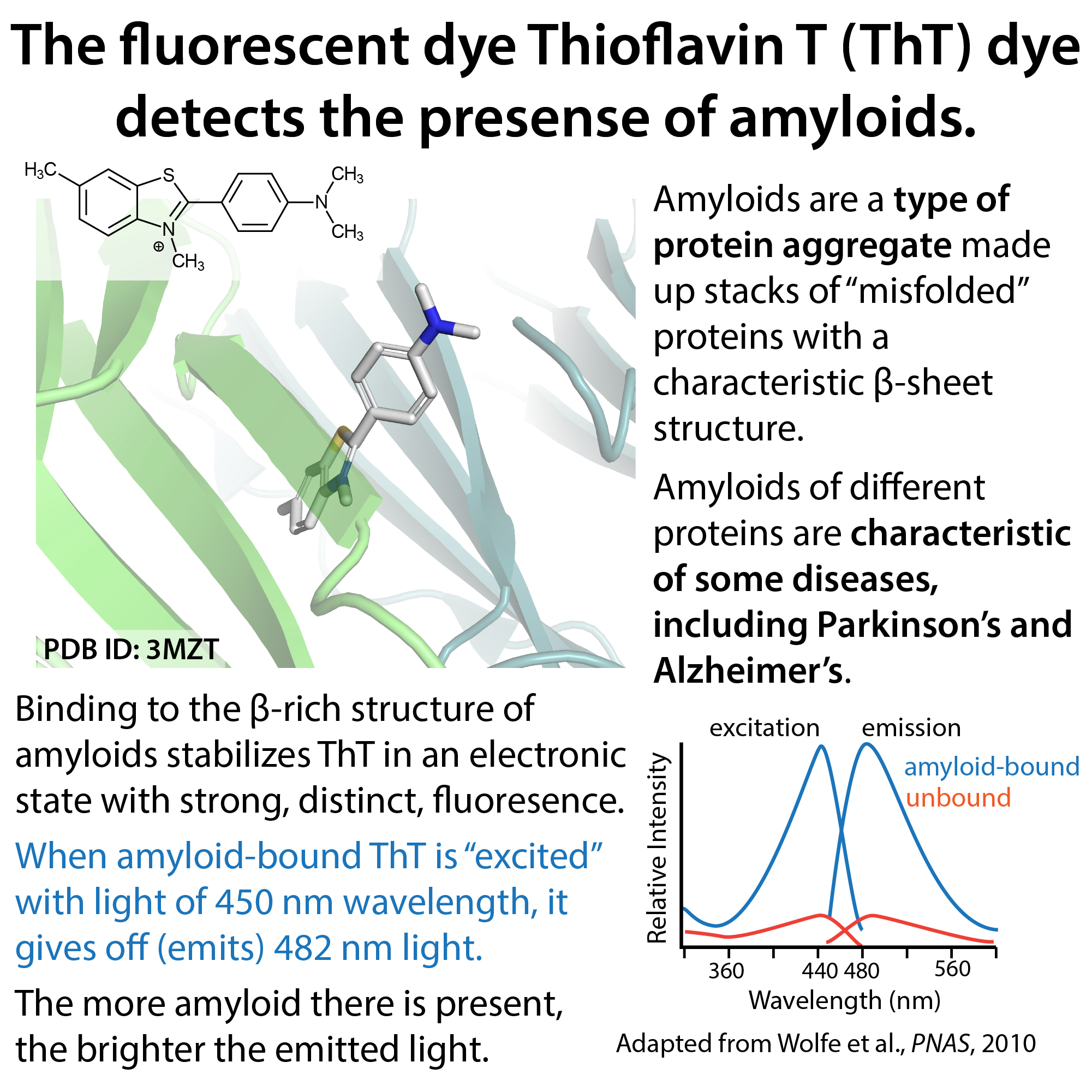
Use of fluorescent dyes greatly sped up the process! Thioflavin T (ThT) binds to the β-rich structure of amyloids (of “any” proteins) and fluoresces (gives off light of a distinct wavelength when light of another distinct wavelength is shone on it). Although ThT can fluoresce even when not bound, the fluorescence is greatly enhanced when bound to amyloids, and the wavelength of light it gives off is shifted, allowing for the direct correlation between fluorescent signal and amyloid concentration (although the correlation factor will depend in part on the structure of the aggregates, which affect how the dye binds). Now, the assay, though not trivial, can be run in a high-throughput (lots of samples at a time) in large-iPhone-sized multi-well plates (think miniature muffin pans), with fluorescence measured in bench-top machines (at least some of the protocols).

There still are barriers to implementation at the clinical level, however. Chiefly, sample obtainment, given the localization of neurodegenerative disease-associated amyloids in the nervous system. Early research using these assays (and much current biomedical research) used samples from donated cadaver tissue. It was later found that misfolded proteins could also be found in CSF (cerebrospinal fluid). Much better than needing to be dead, but still not ideal, as it requires a lumbar puncture (spinal tap). Some scientists, therefore, are studying whether alternative sources such as skin biopsies (containing the endings of nerve cells) might contain enough of the misfolded proteins to be useful biomarker-wise. Perhaps, with enough optimization, even less invasive samples (e.g. blood) could be used (it has been done (see Kluge et al., though according to what I’ve read (see Kuang et al.) that’s hindered by both low concentration and the presence of things like lipoproteins that interfere with the assay). . .
Even if CSF is required, however, SAAs could serve as a great diagnostic and clinical research tool. One reason is its extremely sensitive nature. The misfolded proteins tend to be at very low levels, making them hard to detect with methods such as antibodies (which also might not recognize modified or slightly-differently-folded versions of the proteins). SAAs, however, amplify the original signal dramatically, allowing even low quantities (a single aggregate!) to be reliably measured.
Another reason for SAAs utility is its ability to distinguish between different disease-specific strains/polymorphs of the “same” protein. For example, scientists found that, based on the kinetics of fibril production (how fast the fibrils formed) determined by SAAs, they could differentiate between samples of patients with PD and those with a related but more serious condition called Multiple System Atrophy (MSA) even though both conditions involve the formation of α-synuclein amyloids. In case you were wondering, they found that MSA fibrils form faster but reach a lower maximum fluorescence.
Furthermore, they found that aggregates could even be found in patients pre-diagnosis (with early, “prodromal” symptoms such as sleep disturbances who later went on to develop PD).
SAA tests are also being tested for use in other amyloid-associated diseases including AD, frontotemporal dementia, even chronic traumatic brain injury! Here’s hoping that they can help give scientists and clinicians a heads-start on stopping these diseases. And help scientists figure out how to stop them once detected. These are still early days of SAAs, so I expect further advances are yet to come (especially regarding automation, standardization, true quantification (made difficult by factors like differences in how dyes interact with different aggregates), and compatibility with different tissue types). Definitely an area to watch and I wish all involved the best! Thank you for your work to help people like my dad.
Bibliography
- Development of SAAs
- Concha-Marambio, L.; Pritzkow, S.; Shahnawaz, M.; Farris, C. M.; Soto, C. Seed Amplification Assay for the Detection of Pathologic Alpha-Synuclein Aggregates in Cerebrospinal Fluid. Nat Protoc 2023, 18 (4), 1179–1196. https://doi.org/10.1038/s41596-022-00787-3.
- The intro of this does a great job reviewing the history of the development of SAAs in general, highlighting key papers and milestones.
- Kuang, Y.; Mao, H.; Huang, X.; Chen, M.; Dai, W.; Gan, T.; Wang, J.; Sun, H.; Lin, H.; Liu, Q.; Yang, X.; Xu, P.-Y. α-Synuclein Seeding Amplification Assays for Diagnosing Synucleinopathies: An Innovative Tool in Clinical Implementation. Transl Neurodegener 2024, 13, 56. https://doi.org/10.1186/s40035-024-00449-2.
- Old-school original
- Kocisko, D. A.; Come, J. H.; Priola, S. A.; Chesebro, B.; Raymond, G. J.; Lansbury, P. T.; Caughey, B. Cell-Free Formation of Protease-Resistant Prion Protein. Nature 1994, 370 (6489), 471–474. https://doi.org/10.1038/370471a0.
- Original SMCA
- Saborio, G. P.; Permanne, B.; Soto, C. Sensitive Detection of Pathological Prion Protein by Cyclic Amplification of Protein Misfolding. Nature 2001, 411 (6839), 810–813. https://doi.org/10.1038/35081095.
- Saá, P.; Castilla, J.; Soto, C. Ultra-Efficient Replication of Infectious Prions by Automated Protein Misfolding Cyclic Amplification. Journal of Biological Chemistry 2006, 281 (46), 35245–35252. https://doi.org/10.1074/jbc.M603964200.
- Here, they were able to automate things
- Original RT-QuIC
- Atarashi, R.; Wilham, J. M.; Christensen, L.; Hughson, A. G.; Moore, R. A.; Johnson, L. M.; Onwubiko, H. A.; Priola, S. A.; Caughey, B. Simplified Ultrasensitive Prion Detection by Recombinant PrP Conversion with Shaking. Nat Methods 2008, 5 (3), 211–212. https://doi.org/10.1038/nmeth0308-211.
- Atarashi, R.; Satoh, K.; Sano, K.; Fuse, T.; Yamaguchi, N.; Ishibashi, D.; Matsubara, T.; Nakagaki, T.; Yamanaka, H.; Shirabe, S.; Yamada, M.; Mizusawa, H.; Kitamoto, T.; Klug, G.; McGlade, A.; Collins, S. J.; Nishida, N. Ultrasensitive Human Prion Detection in Cerebrospinal Fluid by Real-Time Quaking-Induced Conversion. Nat Med 2011, 17 (2), 175–178. https://doi.org/10.1038/nm.2294.
- Here’s where they introduced periodic fluorescence measurements (the “real-time” aspect
- Strains
- Summary article (short, more accessible to a broader audience)
- Gerez, J. A.; Riek, R. Neurodegenerative Diseases Distinguished through Protein-Structure Analysis. Nature 2020, 578 (7794), 223–224. https://doi.org/10.1038/d41586-020-00131-3.
- Review article:
- Malfertheiner, K.; Stefanova, N.; Heras-Garvin, A. The Concept of α-Synuclein Strains and How Different Conformations May Explain Distinct Neurodegenerative Disorders. Front. Neurol. 2021, 12. https://doi.org/10.3389/fneur.2021.737195.
- Research article:
- Shahnawaz, M.; Mukherjee, A.; Pritzkow, S.; Mendez, N.; Rabadia, P.; Liu, X.; Hu, B.; Schmeichel, A.; Singer, W.; Wu, G.; Tsai, A.-L.; Shirani, H.; Nilsson, K. P. R.; Low, P. A.; Soto, C. Discriminating α-Synuclein Strains in Parkinson’s Disease and Multiple System Atrophy. Nature 2020, 578 (7794), 273–277. https://doi.org/10.1038/s41586-020-1984-7.
- Here, they show that there’s lower ThT fluorescence with MSA compared to PD, but faster aggregation times. They also use alternative methods to explore structural differences between α-syn aggregates in MSA and PD.
- Shahnawaz, M.; Mukherjee, A.; Pritzkow, S.; Mendez, N.; Rabadia, P.; Liu, X.; Hu, B.; Schmeichel, A.; Singer, W.; Wu, G.; Tsai, A.-L.; Shirani, H.; Nilsson, K. P. R.; Low, P. A.; Soto, C. Discriminating α-Synuclein Strains in Parkinson’s Disease and Multiple System Atrophy. Nature 2020, 578 (7794), 273–277. https://doi.org/10.1038/s41586-020-1984-7.
- Summary article (short, more accessible to a broader audience)
- Potential use of SAAs for diagnostics (mostly PD)
- Reviews
- Calabresi, P.; Mechelli, A.; Natale, G.; Volpicelli-Daley, L.; Di Lazzaro, G.; Ghiglieri, V. Alpha-Synuclein in Parkinson’s Disease and Other Synucleinopathies: From Overt Neurodegeneration Back to Early Synaptic Dysfunction. Cell Death Dis 2023, 14 (3), 176. https://doi.org/10.1038/s41419-023-05672-9.
- Coysh, T.; Mead, S. The Future of Seed Amplification Assays and Clinical Trials. Front. Aging Neurosci. 2022, 14. https://doi.org/10.3389/fnagi.2022.872629.
- This one talks about use of SAAs for a variety of diseases
- Magalhães, P.; Lashuel, H. A. Opportunities and Challenges of Alpha-Synuclein as a Potential Biomarker for Parkinson’s Disease and Other Synucleinopathies. npj Parkinsons Dis. 2022, 8 (1), 93. https://doi.org/10.1038/s41531-022-00357-0.
- Oliveira, L. M. A.; Gasser, T.; Edwards, R.; Zweckstetter, M.; Melki, R.; Stefanis, L.; Lashuel, H. A.; Sulzer, D.; Vekrellis, K.; Halliday, G. M.; Tomlinson, J. J.; Schlossmacher, M.; Jensen, P. H.; Schulze-Hentrich, J.; Riess, O.; Hirst, W. D.; El-Agnaf, O.; Mollenhauer, B.; Lansbury, P.; Outeiro, T. F. Alpha-Synuclein Research: Defining Strategic Moves in the Battle against Parkinson’s Disease. npj Parkinsons Dis. 2021, 7 (1), 65. https://doi.org/10.1038/s41531-021-00203-9.
- This one is broader, but it does a nice job of explaining some of the yet-unanswered questions and challenges with measuring α-syn
- Research
- Here they directly compared methods and protocols carried out in different, independent labs to show reproducibility of results and early diagnostic potential.
- Kang, U. J.; Boehme, A. K.; Fairfoul, G.; Shahnawaz, M.; Ma, T. C.; Hutten, S. J.; Green, A.; Soto, C. Comparative Study of Cerebrospinal Fluid α-Synuclein Seeding Aggregation Assays for Diagnosis of Parkinson’s Disease. Movement Disorders 2019, 34 (4), 536–544. https://doi.org/10.1002/mds.27646.
- Russo, M. J.; Orru, C. D.; Concha-Marambio, L.; Giaisi, S.; Groveman, B. R.; Farris, C. M.; Holguin, B.; Hughson, A. G.; LaFontant, D.-E.; Caspell-Garcia, C.; Coffey, C. S.; Mollon, J.; Hutten, S. J.; Merchant, K.; Heym, R. G.; Soto, C.; Caughey, B.; Kang, U. J. High Diagnostic Performance of Independent Alpha-Synuclein Seed Amplification Assays for Detection of Early Parkinson’s Disease. acta neuropathol commun 2021, 9 (1), 179. https://doi.org/10.1186/s40478-021-01282-8.
- Wang, Z.; Becker, K.; Donadio, V.; Siedlak, S.; Yuan, J.; Rezaee, M.; Incensi, A.; Kuzkina, A.; Orrú, C. D.; Tatsuoka, C.; Liguori, R.; Gunzler, S. A.; Caughey, B.; Jimenez-Capdeville, M. E.; Zhu, X.; Doppler, K.; Cui, L.; Chen, S. G.; Ma, J.; Zou, W.-Q. Skin α-Synuclein Aggregation Seeding Activity as a Novel Biomarker for Parkinson Disease. JAMA Neurology 2021, 78 (1), 30–40. https://doi.org/10.1001/jamaneurol.2020.3311.
- Here’s one where they tested on skin samples.
- Kluge, A.; Schaeffer, E.; Bunk, J.; Sommerauer, M.; Röttgen, S.; Schulte, C.; Roeben, B.; von Thaler, A.-K.; Welzel, J.; Lucius, R.; Heinzel, S.; Xiang, W.; Eschweiler, G. W.; Maetzler, W.; Suenkel, U.; Berg, D. Detecting Misfolded α-Synuclein in Blood Years before the Diagnosis of Parkinson’s Disease. Movement Disorders 2024, 39 (8), 1289–1299. https://doi.org/10.1002/mds.29766.
- Blood!
- Here they directly compared methods and protocols carried out in different, independent labs to show reproducibility of results and early diagnostic potential.
- Reviews
- Thioflavin T (ThT)
- Wolfe, L. S.; Calabrese, M. F.; Nath, A.; Blaho, D. V.; Miranker, A. D.; Xiong, Y. Protein-Induced Photophysical Changes to the Amyloid Indicator Dye Thioflavin T. Proc Natl Acad Sci U S A 2010, 107 (39), 16863–16868. https://doi.org/10.1073/pnas.1002867107.
- Wolfe, L. S.; Calabrese, M. F.; Nath, A.; Blaho, D. V.; Miranker, A. D.; Xiong, Y. Protein-Induced Photophysical Changes to the Amyloid Indicator Dye Thioflavin T. Proc Natl Acad Sci U S A 2010, 107 (39), 16863–16868. https://doi.org/10.1073/pnas.1002867107.
- Concha-Marambio, L.; Pritzkow, S.; Shahnawaz, M.; Farris, C. M.; Soto, C. Seed Amplification Assay for the Detection of Pathologic Alpha-Synuclein Aggregates in Cerebrospinal Fluid. Nat Protoc 2023, 18 (4), 1179–1196. https://doi.org/10.1038/s41596-022-00787-3.