IMFs (InterMolecular Forces) and the hydrophobic effect
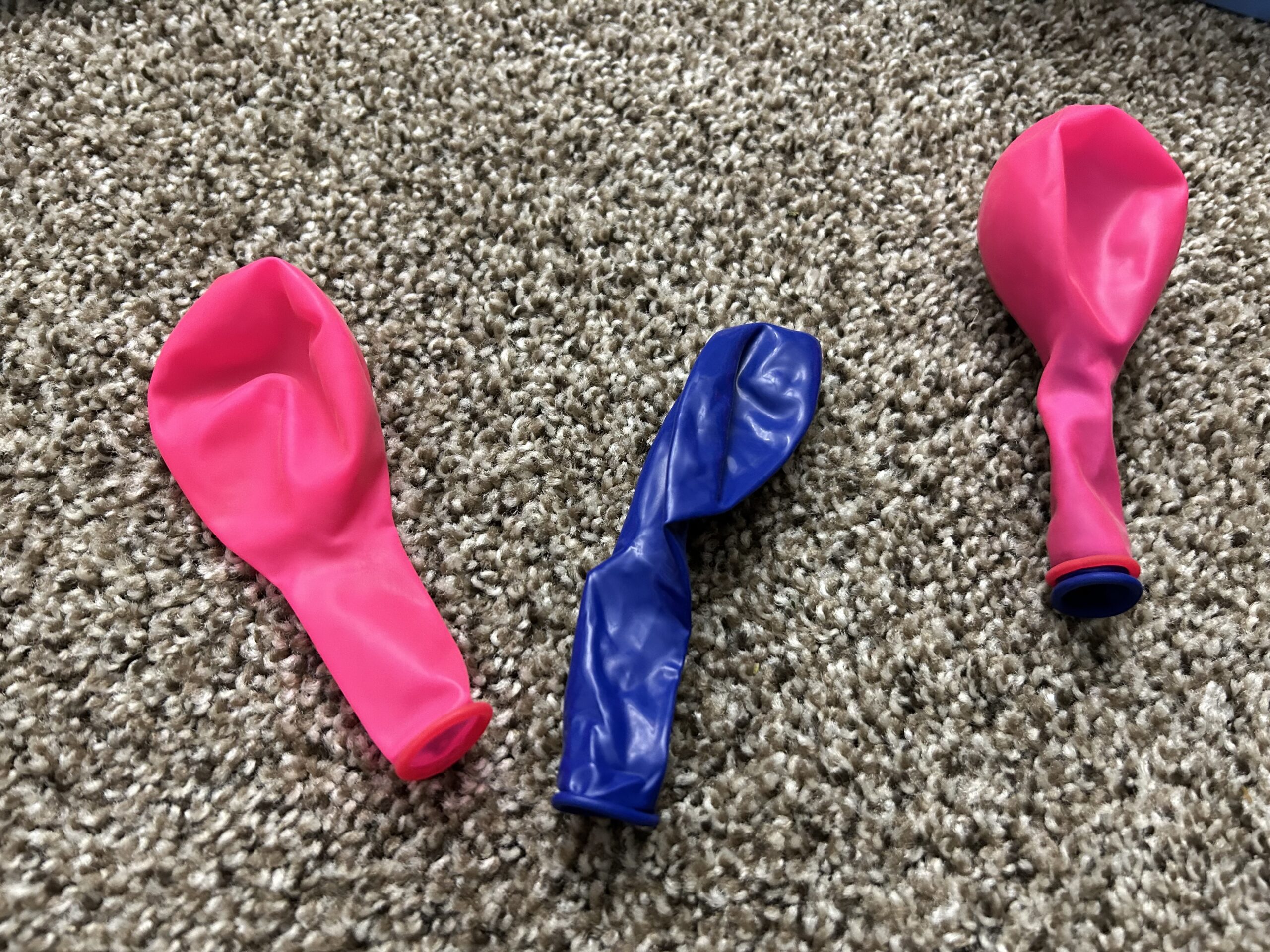
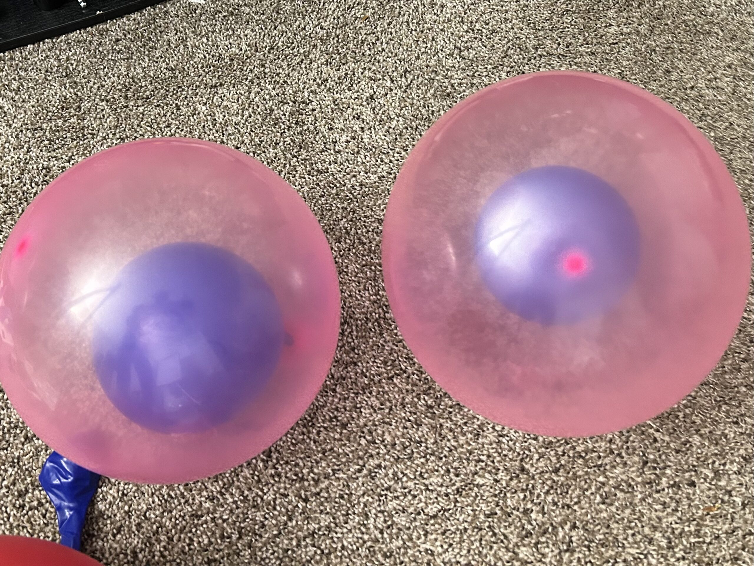
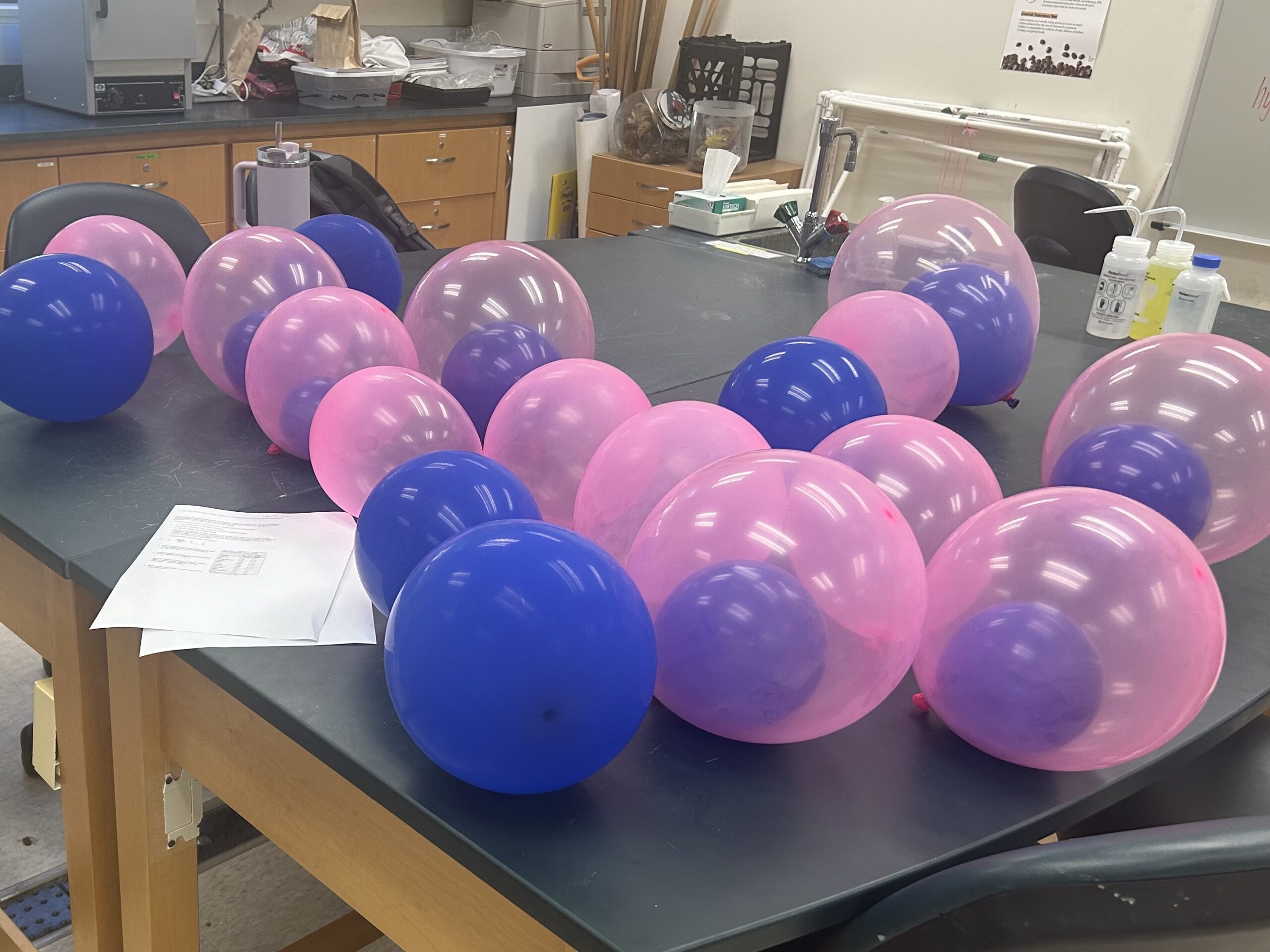
I tried out a balloon dipole demo, and a hydrophobic effect dance today with my students and it was a lot of fun (and hopefully helpful). I started by randomly giving students a blue balloon, a red balloon, 2 blue tied together, 2 red tied together, or a blue inside a red. This started a discussion of how we typically depict negative charge as red and positive as blue. And a review of terms ion, cation, anion, and salt. I had the ions people interact, reinforcing opposites attract, likes repel. And the term “salt bridge”. We also discussed how the larger charges led to higher forces, leading in to a more formal discussion of Coulomb’s law later.
For the balloons inside the balloons, we used them for dipoles – permanent, induced, and random. For the induced, I had students with double balloons interact with ion students and squeeze their balloons to shift the electron density. For the random, I had all the double balloon people next one another, had one of them randomly distribute true electrons to one side and then have the nearby ones get shifted in response. Dispersion forces in action!! I also discussed how if I asked them to spread out randomly around the room, they’d probably evenly space themselves out, but randomness really will have clumps and stuff – explaining why you can flip a coin and get a bunch of tails in a row and how you can get instantaneous dipoles that can set off chain reactions that make for some strong interactions!
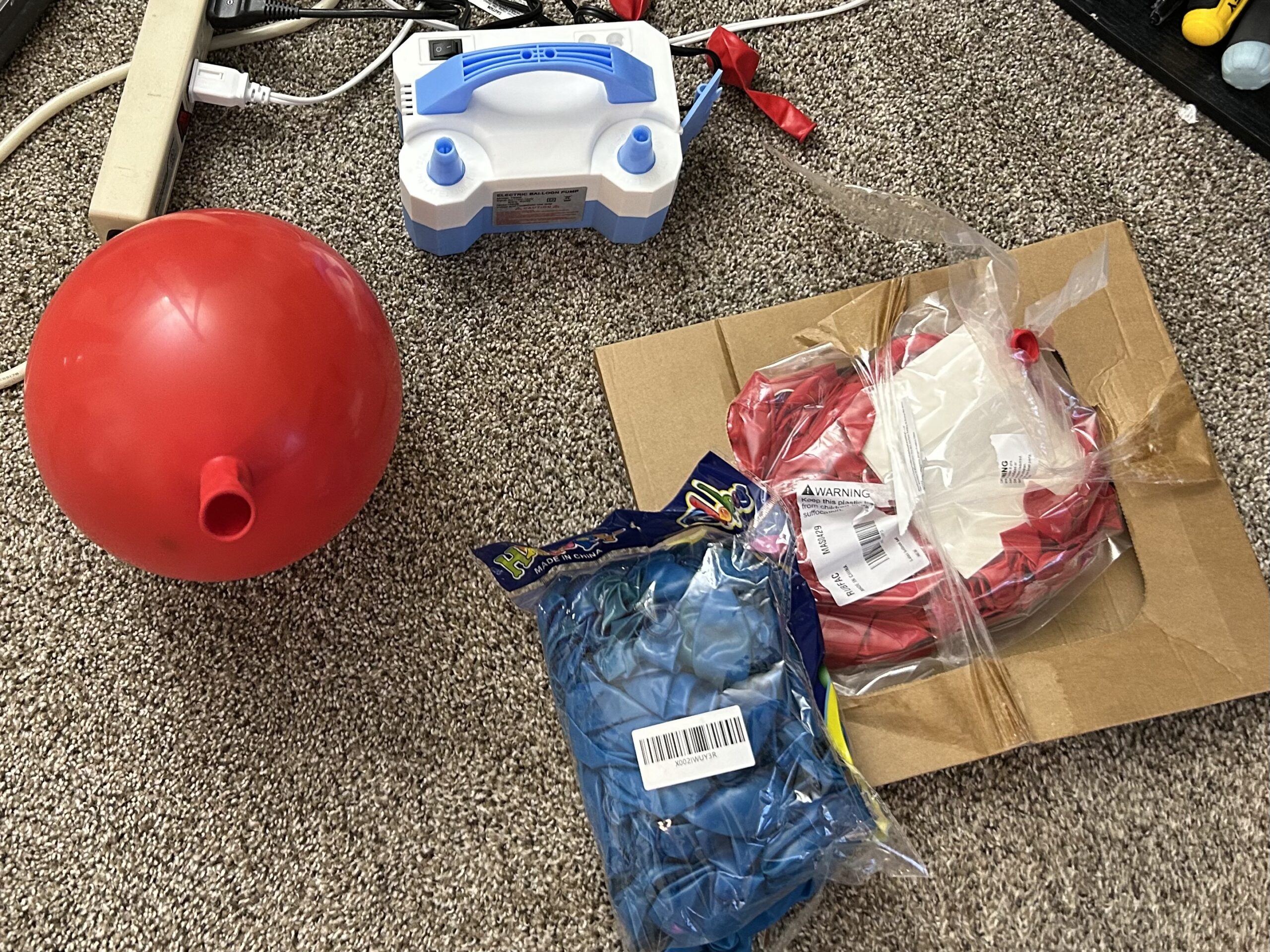
We also acted out the hydrophobic effect. I had most of the students be water molecules, sticking their arms out to serve as hydrogens, and interacting with other people’s oxygen heads. I reinforced the idea of entropy by explaining that because of the ridiculously high molarity of water, they could turn any which way and still find a favorable interaction. So I had them wander around, and pretended to take pictures periodically, discussing how they’d be in different places each time. Entropy!
But then, a student representing a hydrophobic thing came in. And some of the water molecules got stuck orienting themselves around them in order to maximize their favorable interactions. Now, when I “took a picture” those students would be in the same place. Lower entropy!
I had a second student come in and another group of students got tied up. Even lower entropy. But then, I had them force together the hydrophobic things and thus freed up water. Entropy increases!!! Voila – the key basis of the hydrophobic effect. A lot of the students are pre-health so I emphasized how this explains a lot of drug binding (to hydrophobic patches on proteins) but also why so many promising drugs fail for solubility issues.
Cookie Monster acid/base analogy
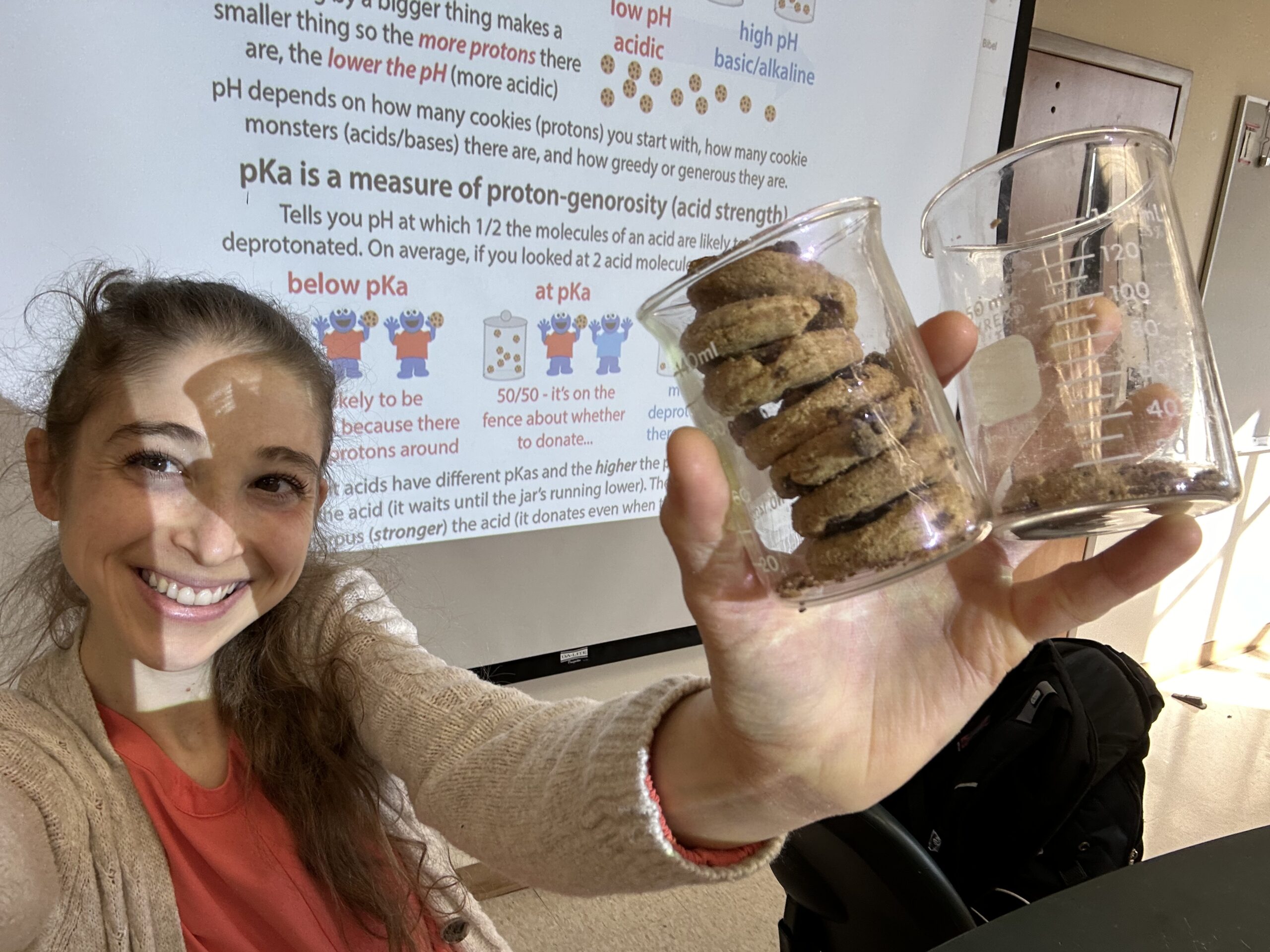
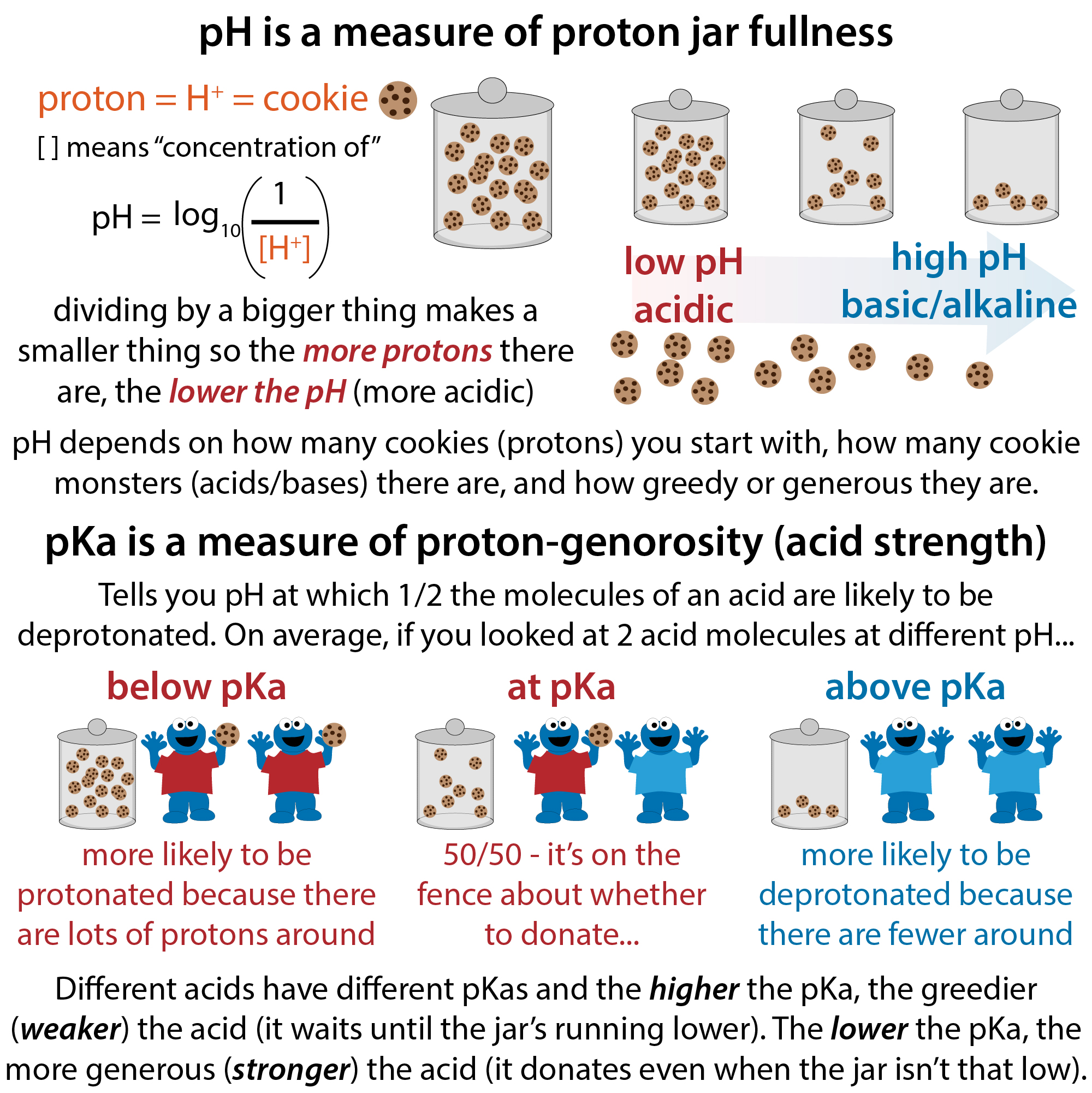
I finally got to try out my Cookie Monster acid/base analogy IRL! Hopefully it softened the blow of having to do acid/base and buffer calculations on the second day of school after summer!
I had beakers filled with different amounts of Famous Amos cookies to represent different pH solutions (fuller cookie jar, lower pH). Then had students pretend to be acids with different pKas and determine when they’d give up their cookies or not. The acid with the lower pKa gave up its cookies unless the jar was super overflowing (low pH) whereas the acid with the higher pKa was stingier and stopped donating at lower levels (higher pH). The one with the lower pKa was thus a stronger acid.
When we talked about buffers and buffering capacity, I compared different concentrations of buffer to having one or two hands to grab or give protons (pretending the hands were separate copies of an acid/base rather than a diprotic one). With fewer hands, we got used up more quickly.
And I had a handy (no pun intended) visual to show the relationship between conjugate acid (cookie in hand) and conjugate base (after giving up cookie).
Protein domain architecture
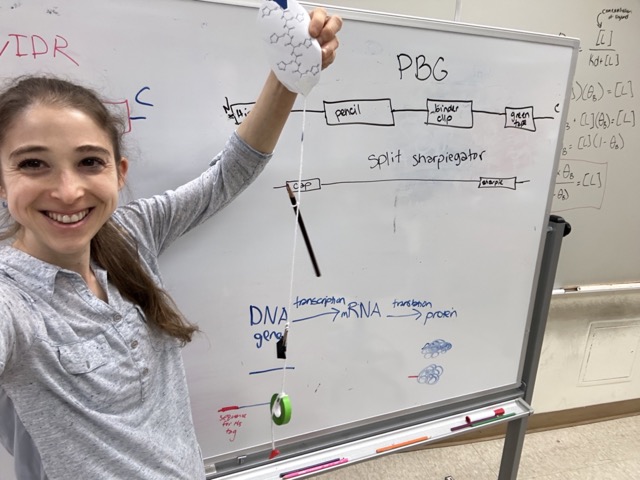
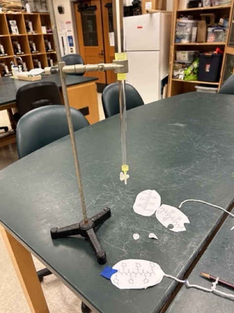
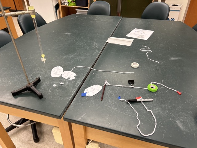
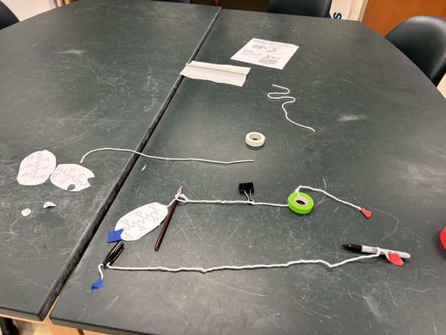
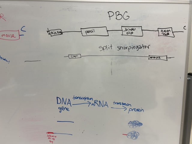
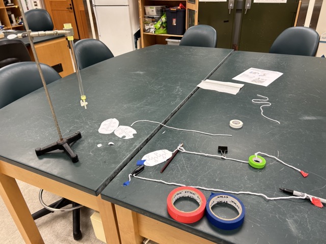
Today was domains day in my biochemistry class. To reinforce our lesson on tertiary and quaternary structure and all the different ways domains can be represented (and the different things the term itself can mean – structural? functional?) I gave students pieces of yarn and had them tie together 3 random objects, which served as “domains.” I had them put blue tape on the N-terminus and red tape on the C-terminus (emphasizing the difference between the actual termini and things that are just “N/C-terminal to…”, then sketch out a domain architecture for their proteins, give them names, and introduce them to us.
We discussed how often (but not always) domains will be distinct structural and functional units like this. And how our proteins could have similar domains – for example, two of us had pencils in our proteins. This led to a discussion of how domains can be evolutionarily conserved, duplicated, etc. or just the same sort of thing stumbled upon twice by evolution.
I also did an example where the functional domain was only formed when parts of the protein that were distant in primary structure (sequence) came together in the tertiary structure – in this case a sharpie and its cap.
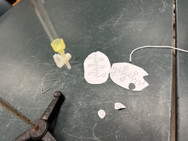
Leading into the technique we will be doing in lab, expressing and purifying His-tagged malate dehydrogenase (MDH), I had the students add a histidine tag (printout of a string of histidines) to their protein – moving the N-terminus in the process. And we discussed how in order to actually get the tag added on, we’d have to add the DNA encoding for it because that’s what we will be sticking in bacteria (Tuesday). I then used my His-tagged protein to walk them through the idea of Ni-affinity chromatography, with pieces of imidazole as competitor.
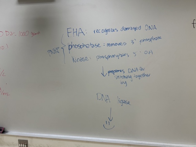
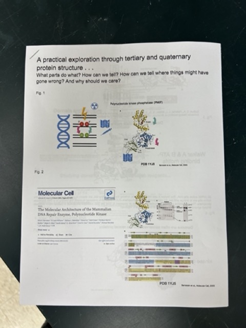
I then discussed the idea of limited proteolysis by talking about how it would be easier to cut the linker regions (yarn) than the domains, and we looked at an example of limited proteolysis in a paper analyzing a protein called PNKP (polynucleotide kinase phosphatase) which gets broken DNA ready for DNA ligase to stitch up. It has 3 domains – an FHA domain that recognizes the site of damage, a kinase domain that phosphorylates one DNA end, and a phosphatase domain that dephosphorylates the other. (Also a great opportunity to reinforce those key enzyme terms that will show up all over the course).
This led to a discussion whereby I had students come up with ways in which they might be able to tease apart what might be going on in the cell and the protein (e.g. which domain) if they saw that DNA wasn’t getting fixed by PNKP.
Hemoglobin activity
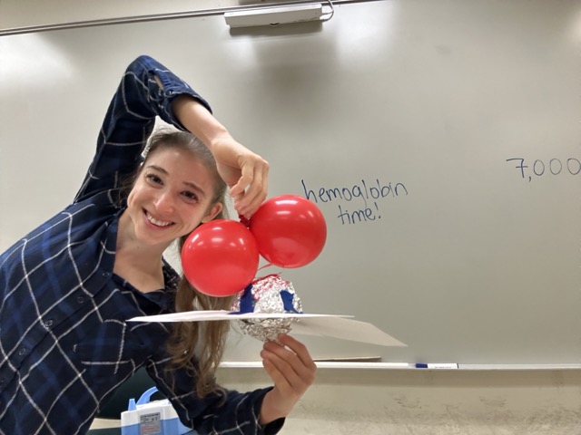
Today was hemoglobin day in my biochemistry class. I had my students act out the structural transitions taking place upon oxygen binding alongside us viewing what was going on in an animation showing structures of hemoglobin in the different forms – love this interactive tutorial from Eric Martz and Frieda Reichsman! http://hemoglobin.molviz.org
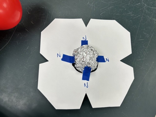
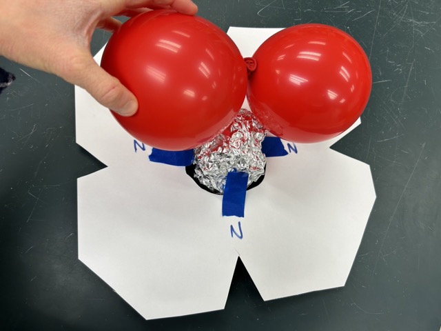
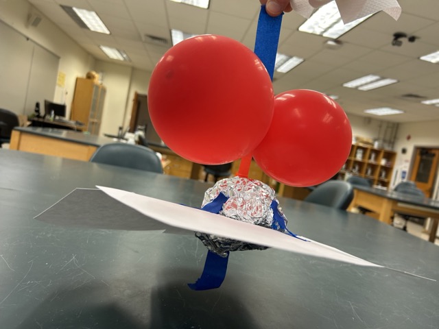
I cut out 4 “hemes” out of cardstock and used 4 pieces of blue tape (nitrogens) to tape a foil ball (iron) into a central hole. Then I had one student hold the heme loosely (for a puckered heme) and another student act as histidines, one binding to the bottom of the iron with a piece of blue tape (another nitrogen interaction). Then, I had that student tape their other hand to a pair of red balloons (oxygens) and tape that to the foil ball (iron), pulling the heme (and their bottom hand) up with it. I emphasized how, in this way, the binding of oxygen causes a conformational change throughout the monomer.
Next, I had the 4 pairs of students come up, unbind the oxygen, and then the 4 that were acting as the histidines link arms (salt bridges), representing the taut deoxygenated conformation. Then I had them try to bind oxygen (with their arms linked) and they realized they’d have to unlink in order to do so. And, once one unlinked, it caused a ripple effect throughout the other subunits, changing them all into the high-affinity state.
Then, we discussed how, although lack of oxygen facilitates the T state, in order to actually form the most effective inter-subunit salt bridges to actually stabilize that state, you had to have the histidine protonated. Cue review of pKr and comparison of the basic amino acids… Hence the Bohr effect whereby low pH (high proton concentration) increases the proportion of histidine that is protonated, allowing for the salt bridges to form and stabilize that form.
Finally, I became 2,3-BPG and crawled under their linked arms to the central cavity (that’s only open in the T state) and discussed how they must be much happier with me there, so 2,3-BPG stabilizes the T state, making it harder for oxygen to bind.
Then, we did some more official stuff with graphs 🙂
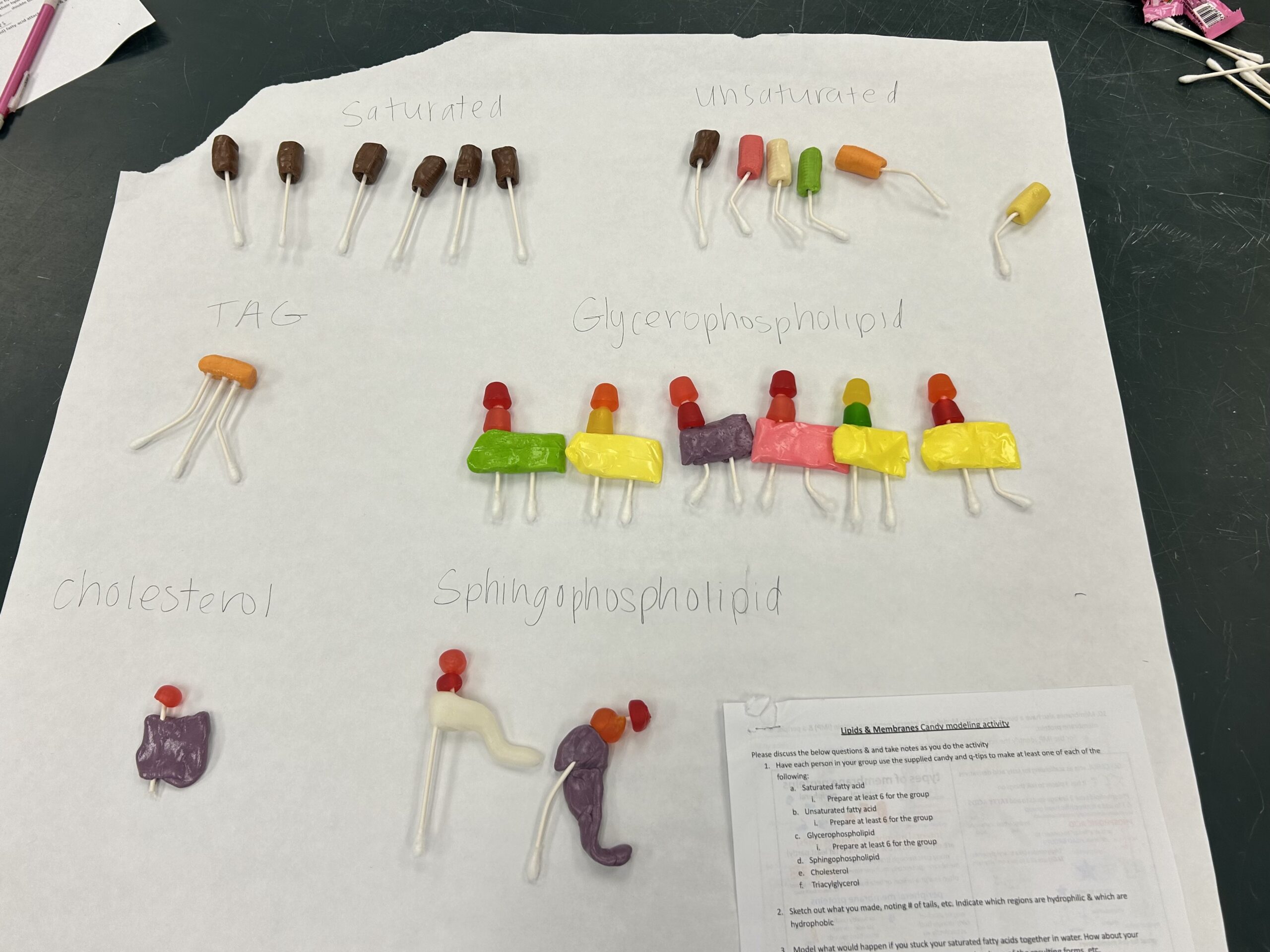
Lipid candy modeling activity
Here are some guided questions I had the students use…
Lipids & Membranes Candy modeling activity
Please discuss the below questions & and take notes as you do the activity
- Have each person in your group use the supplied candy and q-tips to make at least one of each of the following:
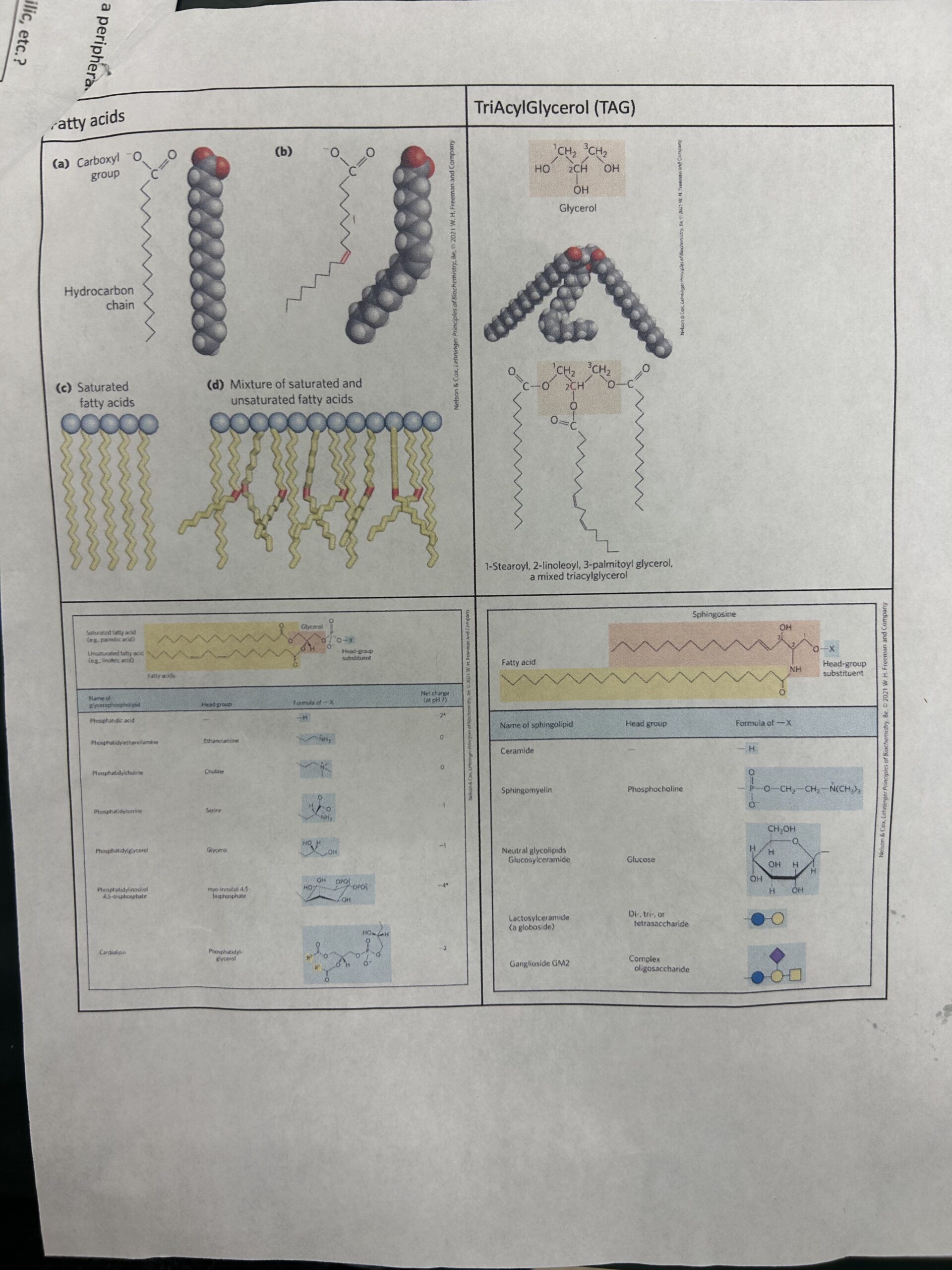
- Saturated fatty acid
- Prepare at least 6 for the group
- Unsaturated fatty acid
- Prepare at least 6 for the group
- Glycerophospholipid
- Prepare at least 6 for the group
- Sphingophospholipid
- Cholesterol
- Triacylglycerol
- Saturated fatty acid
- Sketch out what you made, noting # of tails, etc. Indicate which regions are hydrophilic & which are hydrophobic
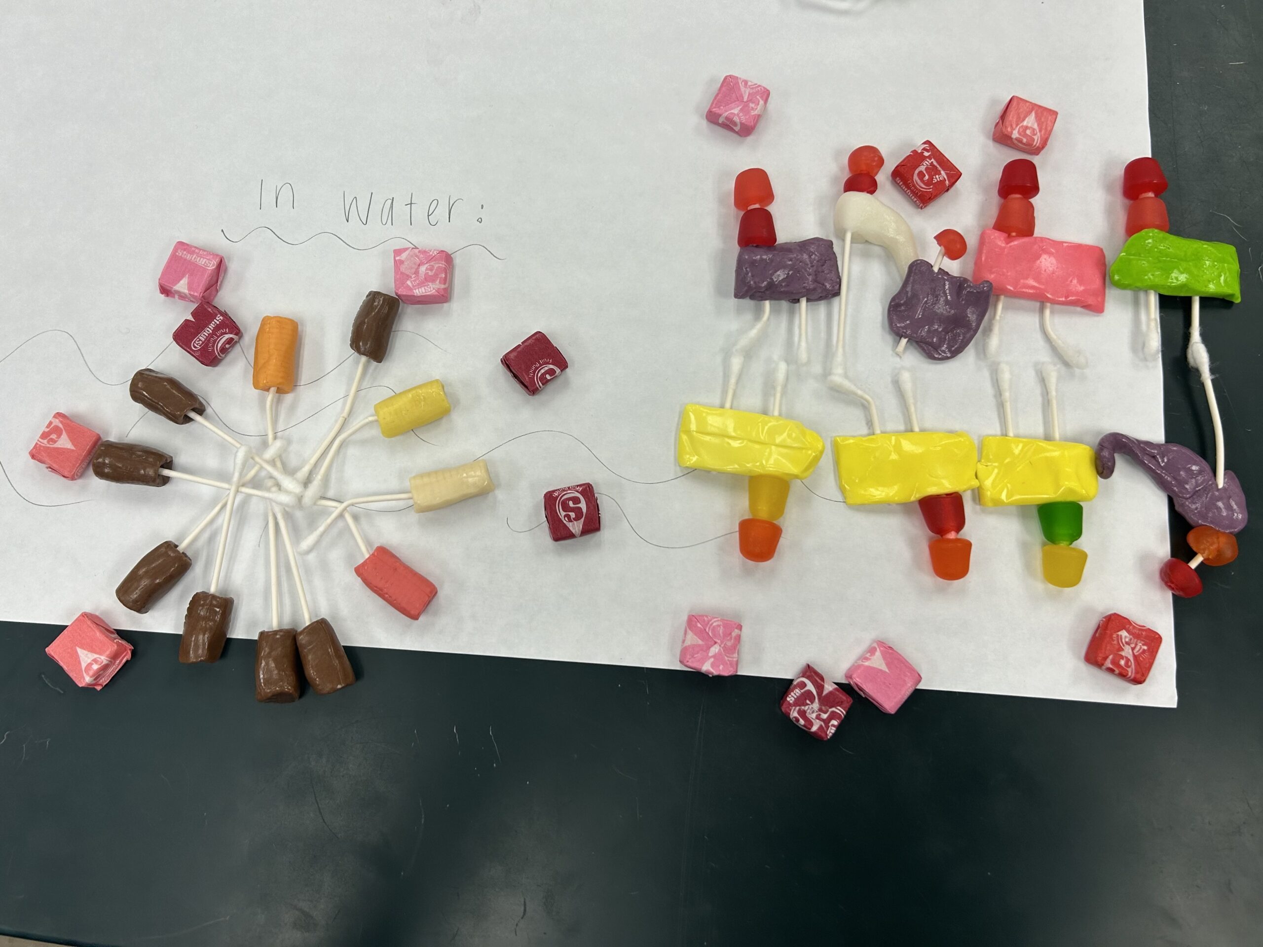
- Model what would happen if you stuck your saturated fatty acids together in water. How about your unsaturated fatty acids? Comment on the packing, the shape of the resulting forms, etc.
- Which would have a higher melting point?
- How could you raise or lower the melting point by altering the composition of fatty acids?
- Model what would happen if you stuck your TAGs together in water. Comment on the packing, the shape of the resulting forms, etc.
- Model what would happen if you stuck your phospholipids (glycero- and sphingo-) together in water. Comment on the packing, the shape of the resulting forms, etc.
- Where could you find modifications and what types of modifications might you find? Go ahead and model some in with your candy.
- Which directions can and can’t the lipids move easily & why?
- How would the membrane look at a higher temperature? A lower temperature?
- Now introduce some cholesterol. Where in your model would it be and how would it be oriented? Does it go all the way through the membrane?
- How would the membrane look at a higher temperature? A lower temperature? How did cholesterol change your answer compared to before?
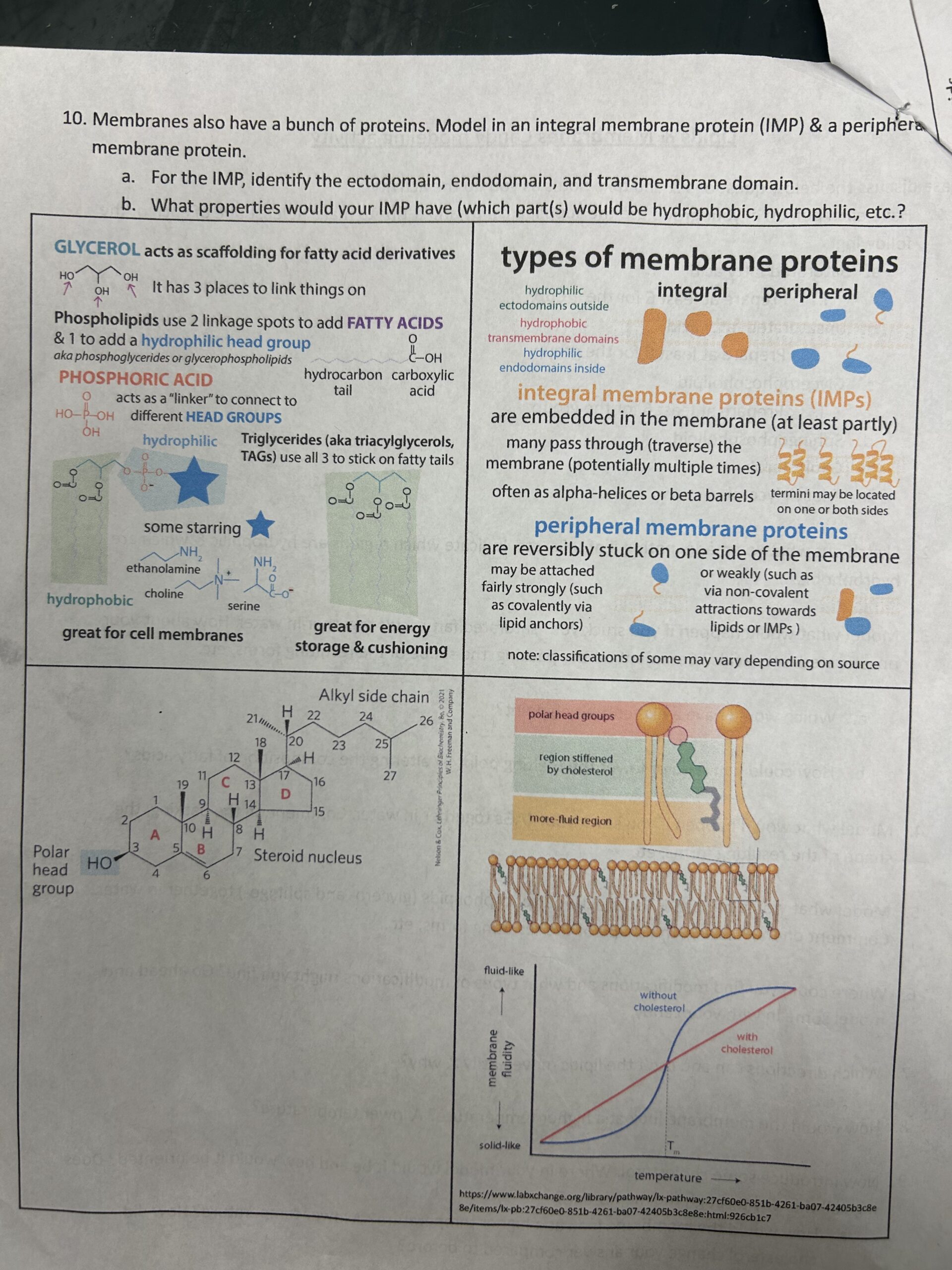
- Membranes also have a bunch of proteins. Model in an integral membrane protein (IMP) & a peripheral membrane protein.
- For the IMP, identify the ectodomain, endodomain, and transmembrane domain.
- What properties would your IMP have (which part(s) would be hydrophobic, hydrophilic, etc.?
and I went ahead and uploaded a PDF to my Google Drive: https://bit.ly/lipid_candy_modeling
MDH m&m’s redox activity
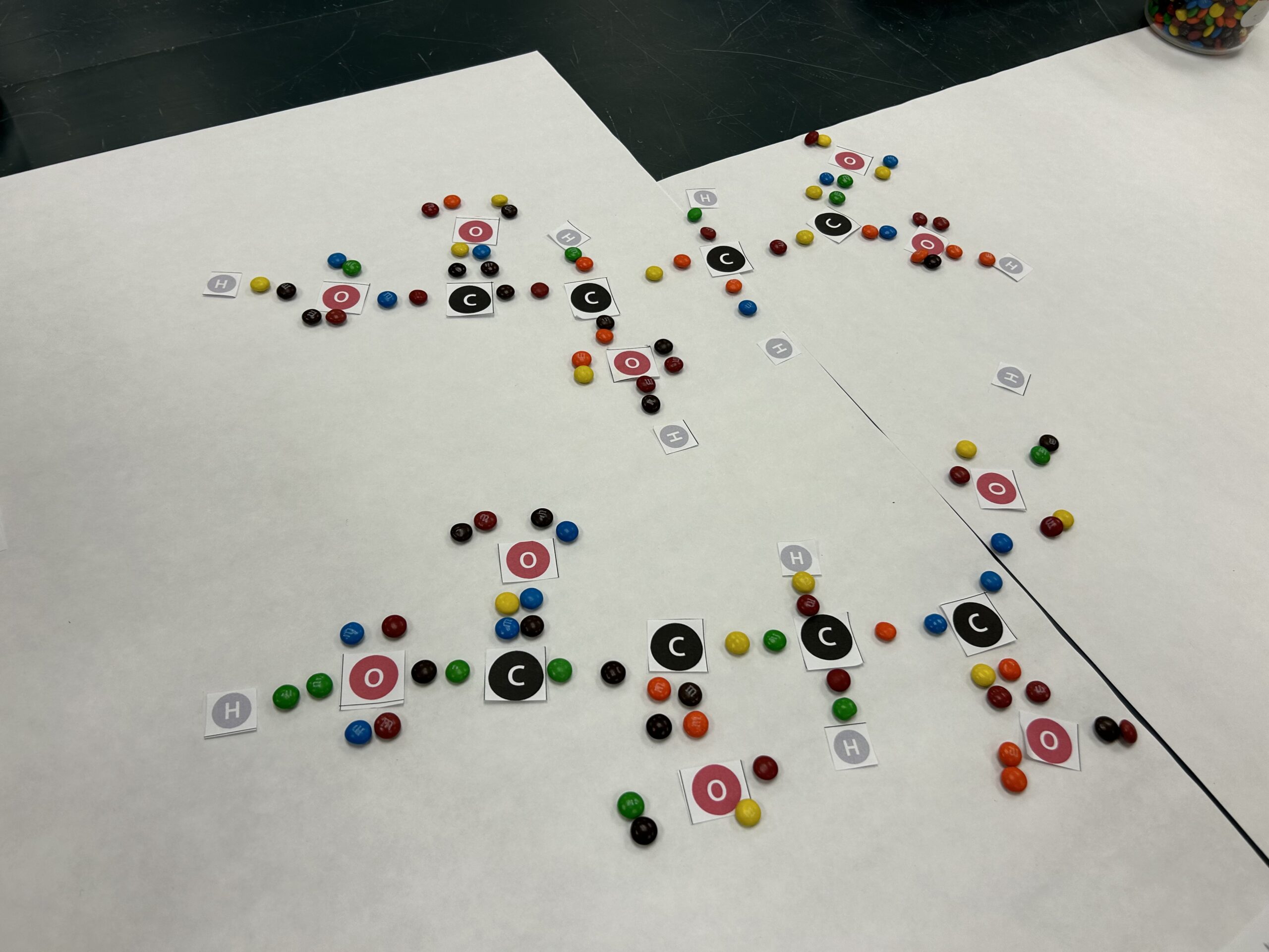
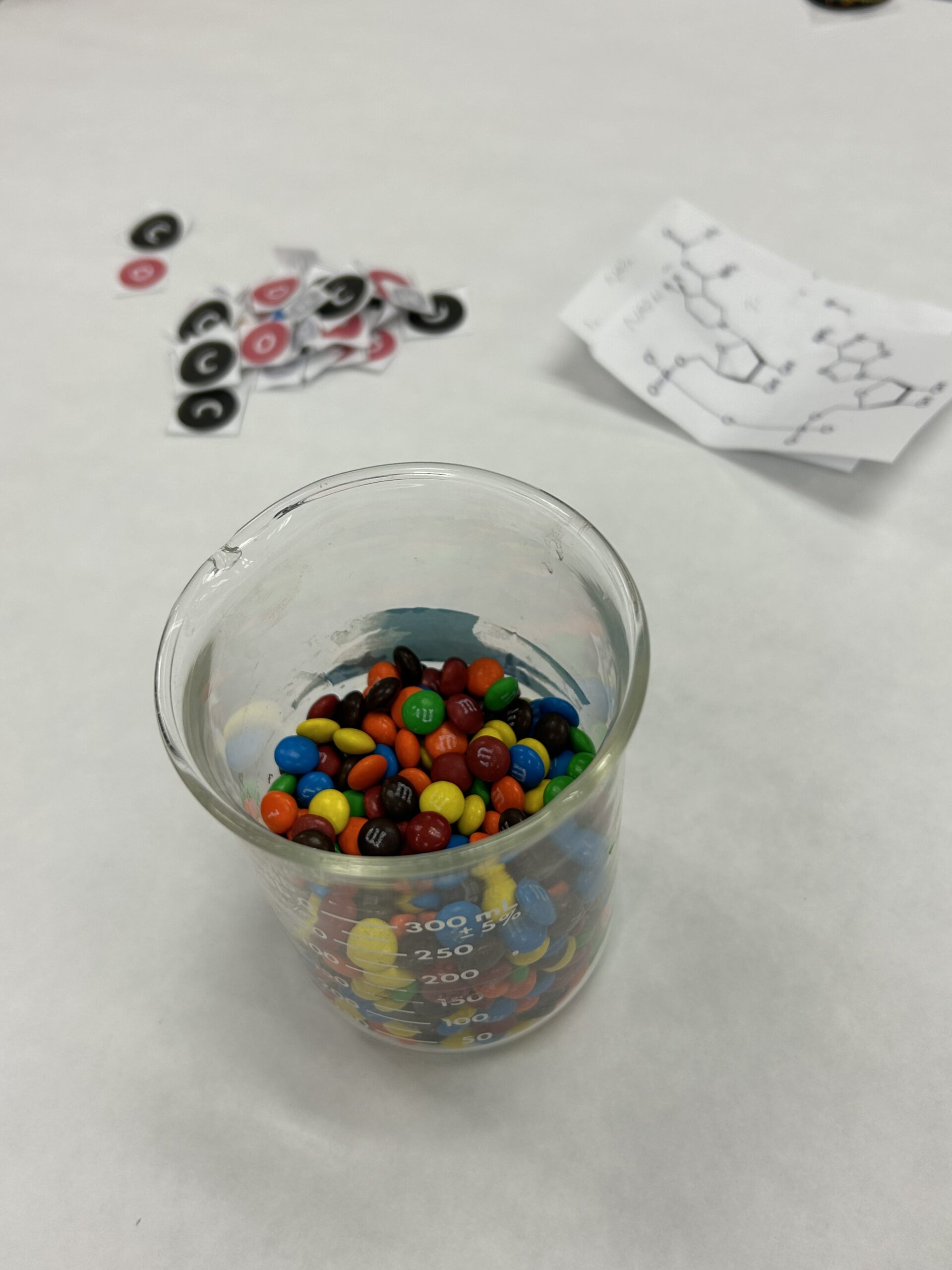
When it comes to redox, the focus is often on the “ox,” but the distinguishing feature of redox is the electrons!!!!!!! To help emphasize this – and get the students engaging on multiple deep levels with the malate dehydrogenase (MDH) reaction, I had them make Lewis structures of malate and oxaloacetate, with mini m&m’s serving as electrons.
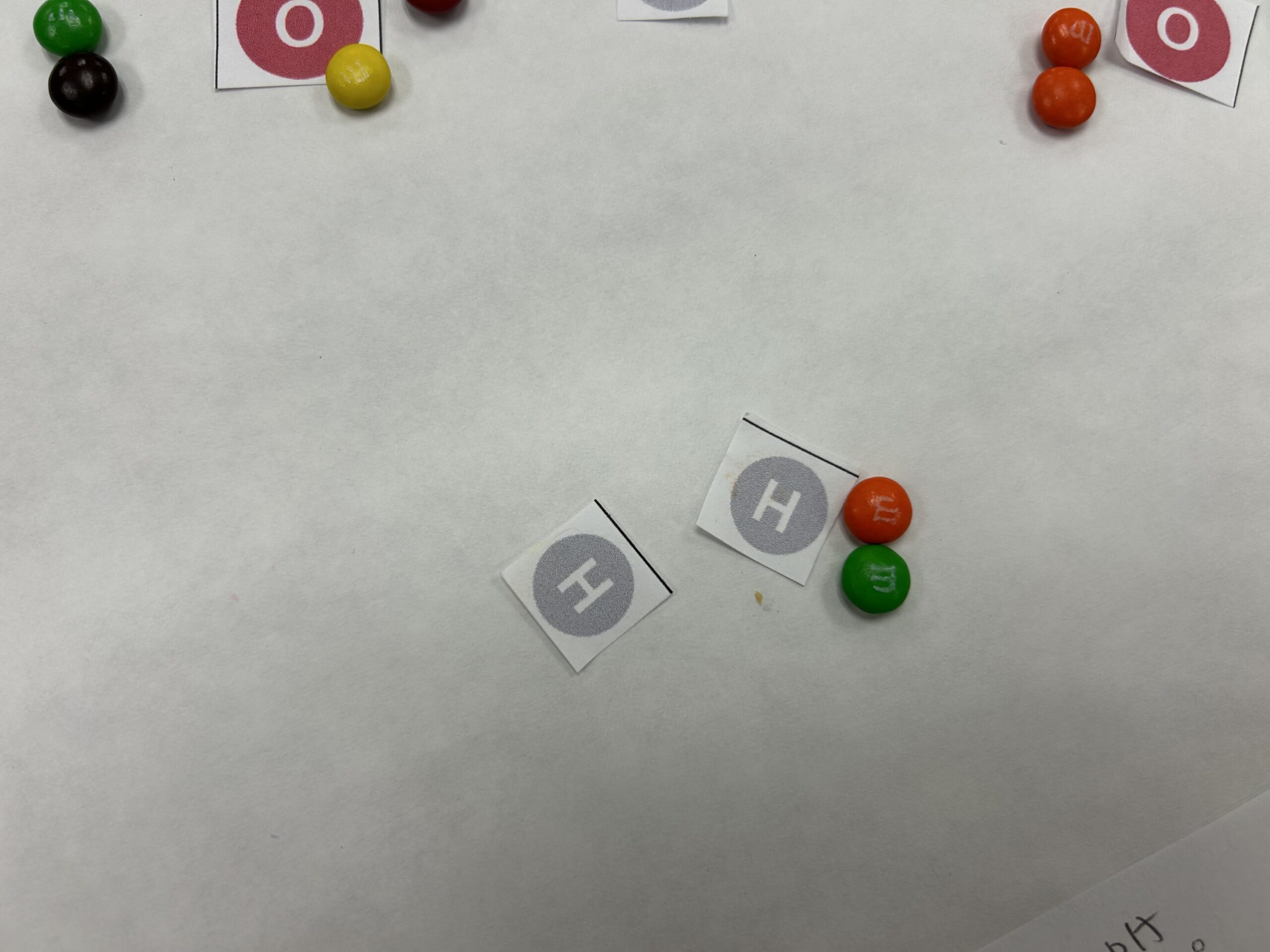
I had them turn their malate into oxaloacetate – which made them see how they had to remove both a proton and a hydride (H with 2 electrons) to do so.
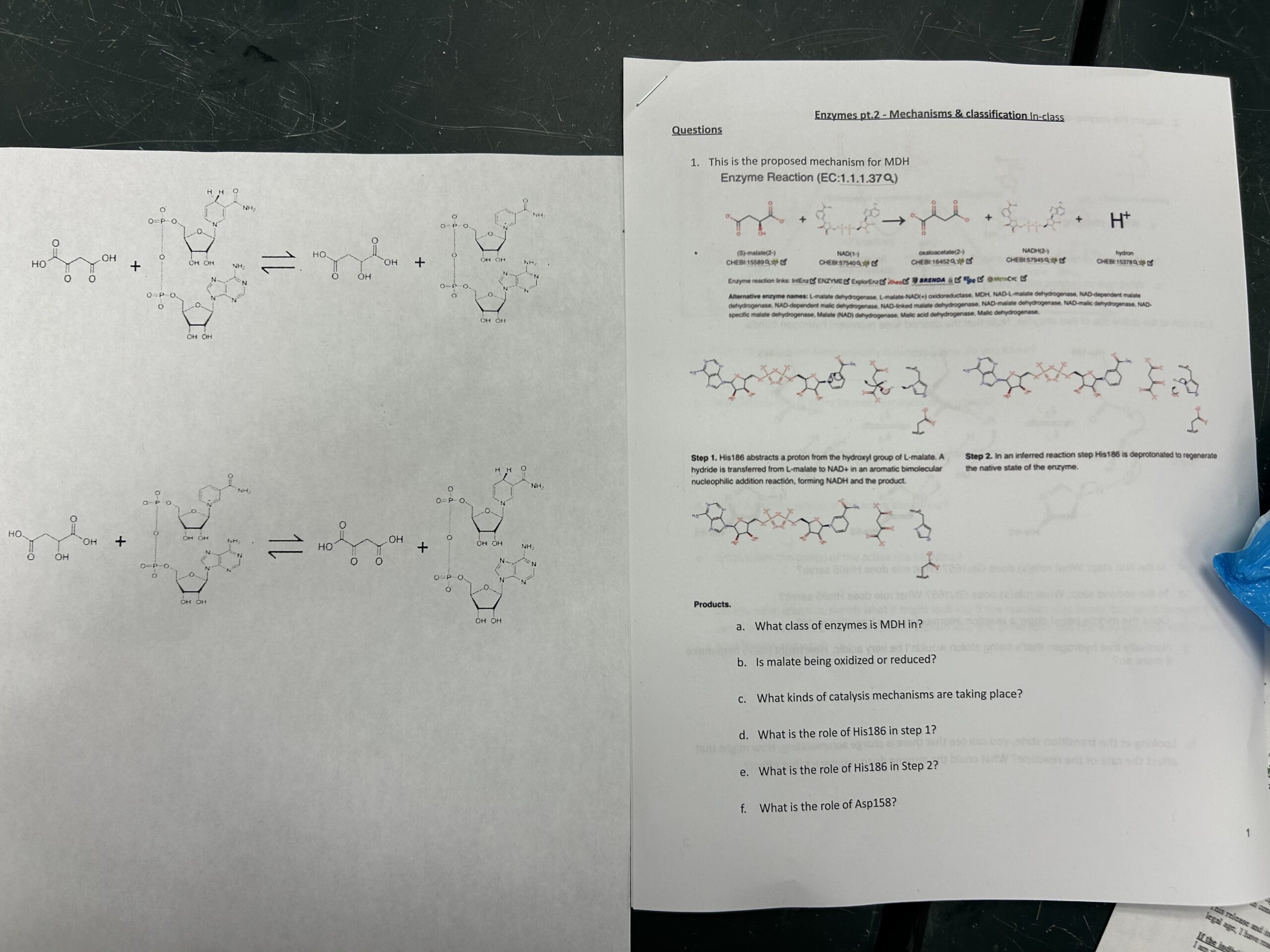
We discussed how those electrons, as part of the hydride, got transferred to NAD+, to give NADH, and we looked at the chemical structures of the two molecules – how the + refers not to an overall positive charge on the molecule (which they saw was negatively-charged overall), but rather to the + charge on the nitrogen. A + charge that allows it to serve as a great “electron sink” for taking those electrons.
We then used the M-CSA (Mechanism and Catalytic Site Atlas) to look at how MDH is able to catalyze this reaction. https://www.ebi.ac.uk/thornton-srv/m-csa/entry/526/ How His acts a general base to steal the proton (helped out by the nearby Asp, with it’s negative charge help making His more grabby (stabilizing the + charge that comes with the proton and thereby raising the pKr). And then the electrons that were attached to the proton push down to make a double bond, kicking out a hydride that attacks the NAD+, leading to the pushing of the ring’s electrons towards the used-to-be-positive N, which would gladly take them.
Then, in order to do it again, the His would have to give up the proton to water to form a hydronium (H3O+) ion (hence the “released” proton).
Then, I had them do the whole malate-to-oxaloacetate thing imagining their hands were the active site of MDH.
And, finally, I had them reverse the reaction, going from oxaloacetate to malate (Which required adding a hydride and proton to oxaloacetate from NADH – thereby depleting NADH (and – as relevant to the afternoon’s lab – the UV340 absorbance!)
Oxidative phosphorylation demo
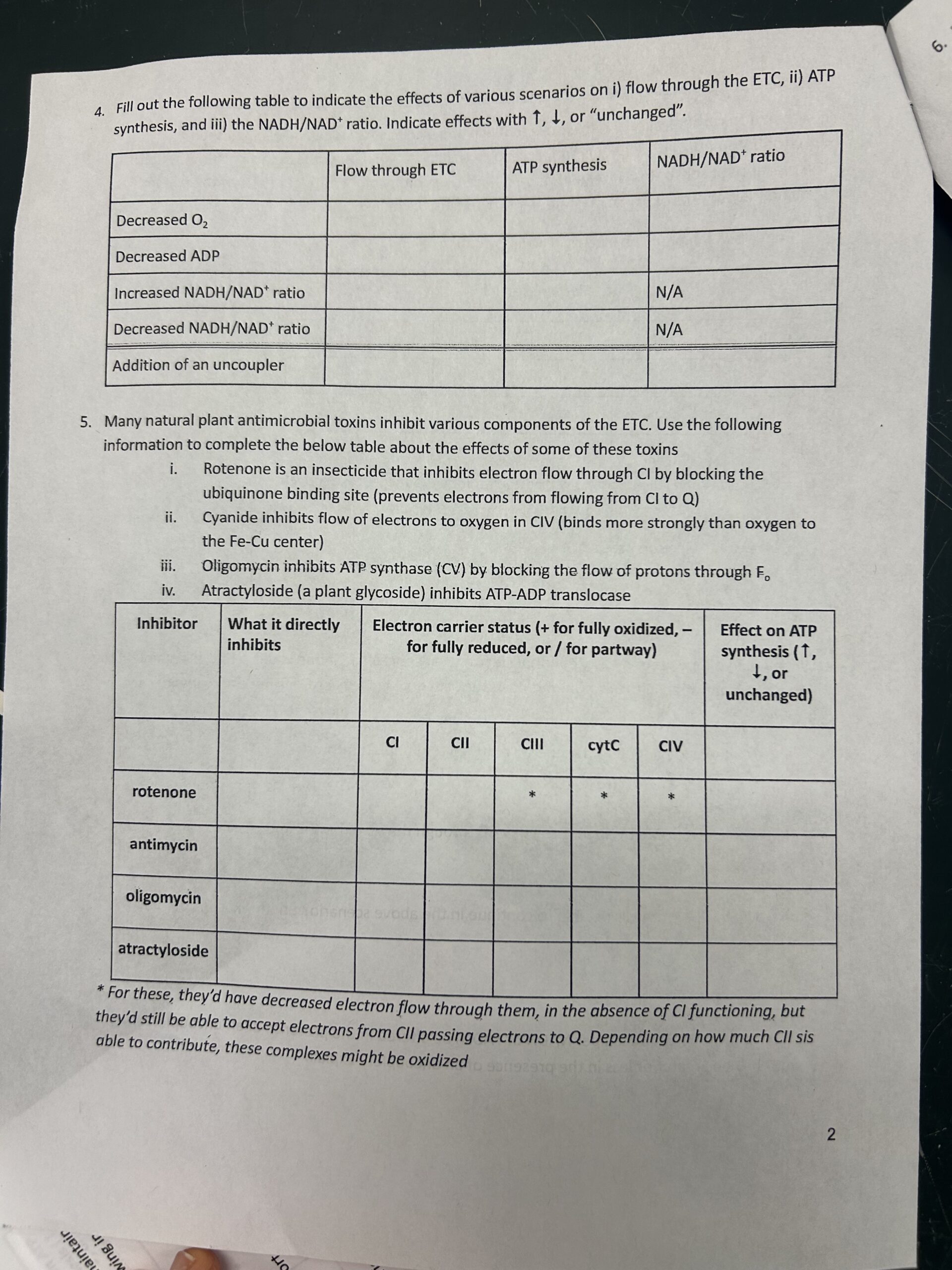
In class today, we covered mitochondrial oxidative phosphorylation. I made little (unlabeled) models of the ETC (electron transport chain) and ATP synthase and had the students label:
- Cytoplasm, intermembrane space, and matrix
- Inner & outer mitochondrial membranes
- Complexes, Q, & cytochrome c
Then, I had them use red mini m&m’s to represent electrons and pass them down the chain, pumping protons (blue m&m’s) out into the intermembrane space as they went (but not at CII because you don’t get enough energy to pump going from FADH2 to Q!), then using the gradient to make ATP and seeing how many ATP they could make if they started with NADH or FAD.
Students expressed that walking through the process tactilely helped them understand it better. And it gave them a model to help them reason through what would happen with regards to ATP production & electron flow in the presence of artificial proton gradients, electron acceptors, uncouplers, and complex inhibitors.
PS – I know that cytochrome c only takes 1 electron at a time, but was simplifying things in the video