I think biochemistry is pretty sweet, but how “sweet”’s your blood after you eat? For people with diabetes, that’s something they might need to measure, and teaching you the science behind it would be my pleasure! The ability of glucose to reduce allows glucometers to deduce, the concentration of sugar after you drink juice, helping diabetics know how much insulin to infuse! But what type of insulin should you use???
Note: this post is adapted from a mashup of past posts with more details I will link to
Insulin is a hormone (chemical messenger) your body uses to help control blood sugar levels. I’m grateful that I don’t have diabetes, but, for people who do, their body has a hard time making and/or “hearing” insulin’s call to “let glucose in!” As a result, they have trouble controlling blood sugar levels, so they often use devices called glucometers to measure blood glucose levels. And then, they may turn to “designer insulin” which can act fast or slow thanks to some cool biochemistry.
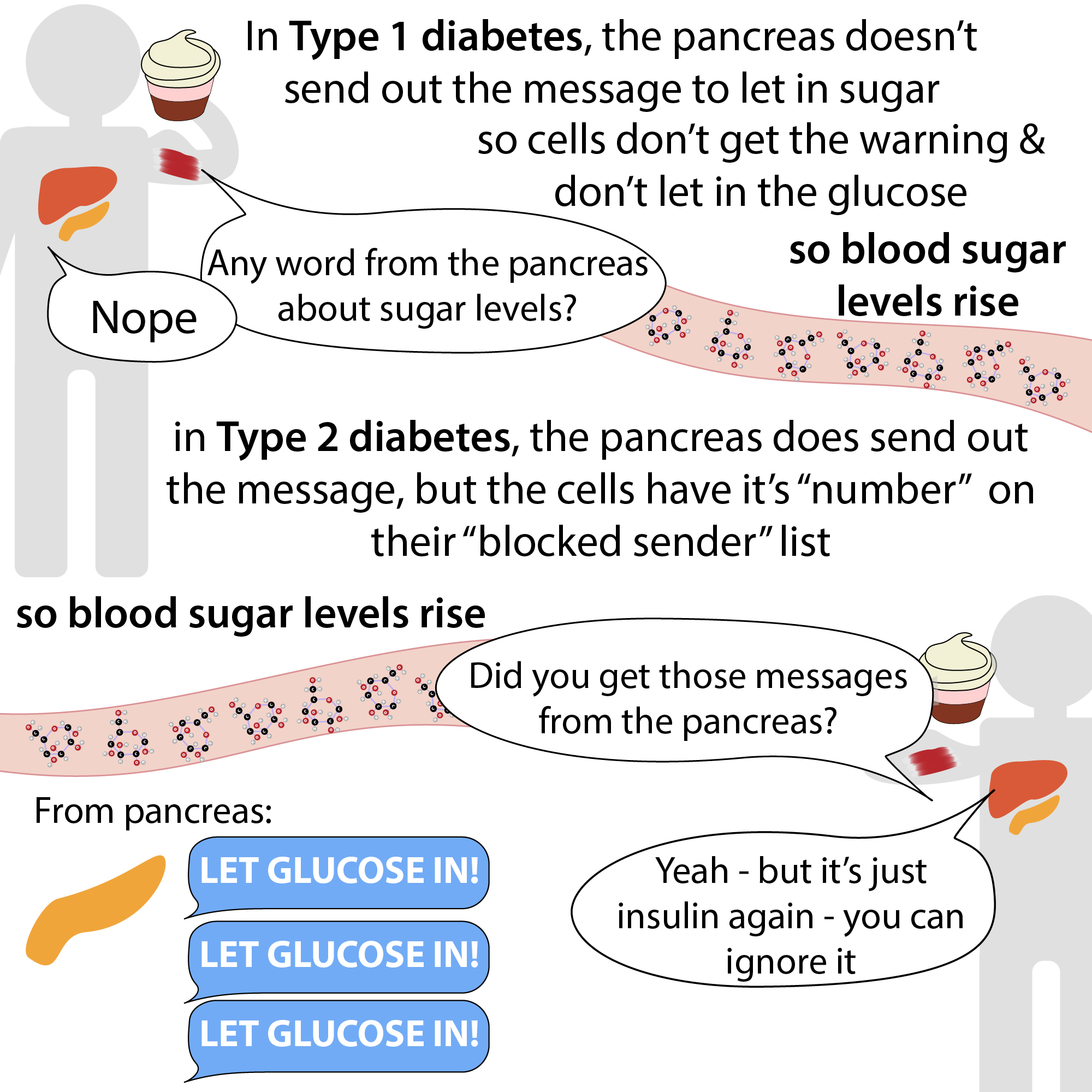
Diabetes is a disease where the body either doesn’t make (type 1 diabetes (T1D)) or can’t use effectively (type 2 diabetes (T2D)) a hormone called insulin. Hormones are chemical messengers that can relay messages throughout & between cells in your body. Different hormones are made of different building blocks & relay different messages.
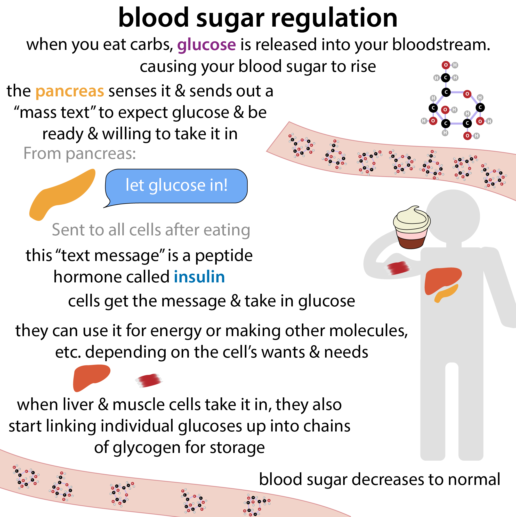
Insulin is a peptide hormone, so it’s made up of amino acids (these are the same “letters” as proteins us but proteins are more like novels while peptide hormones are like “text messages”). The text sender is an organ called the pancreas, & the message it relays is – there’s a lot of sugar in the blood – let’s take some into the cells, use some, & store some, why don’t we?
The pancreas basically sends out a “mass text” to cells throughout the body in the form of insulin. The cells respond by opening their doors to glucose (the main monosaccharide (single sugar unit)), importing it from the bloodstream into the cells thus lowering the amount of glucose in the blood. Since glucose is hydrophilic (water-loving), it needs help getting through the hydrophobic, fatty membranes surrounding a cell. Help is provided via specialized transporter proteins that provide a channel through. These transporters are stored in membrane-bound vesicles inside the cell – when the cell gets insulin’s message (by insulin binding to receptors on the cell surface), those vesicles fuse with the outer membrane, inserting these transporters into it and thus providing a path in.
Additionally, insulin tells cells in the liver to “turn on” an enzyme called glycogen synthase, which strings excess glucose into chains (polysaccharides) called glycogen for storage. more on glycogen here: http://bit.ly/2JwIuRV
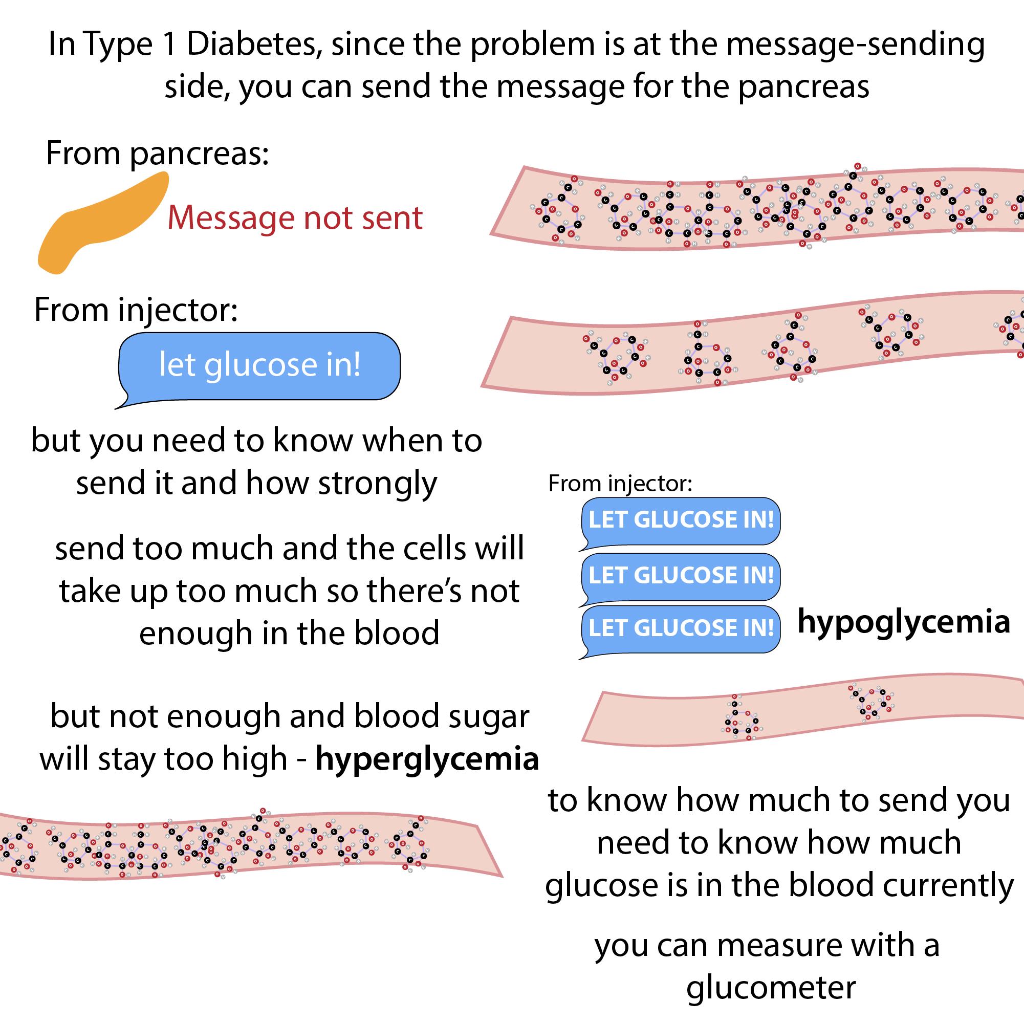
Too much sugar is hypERglycemia (remember ovER). Too little sugar is hyPOglycemia (remember below). Both are bad. Which is why having a properly-functioning “early alert” system through insulin signaling is so important. It gives your cells time to prepare & avert a crisis.
But T1D is like the text message never being sent and T2D is like the text gets sent but the phones have the caller on their block list so they never hear the message. If cells are phones, it’s not that they’re completely turned off – they can still get other messages fine, but their bodies have become “desensitized” to it.
Without a functional natural “early alert” system, diabetics need an “unnatural” way to measure how much glucose is in their blood and, if appropriate, inject/infuse insulin (if sugar’s too high) or eat some sugar if it’s too low. But in order to know what, if any, correcting is required, you need to know what your blood sugar level currently is. And common ways to do this take advantage of glucose’s ability to act as a “reducing agent.”
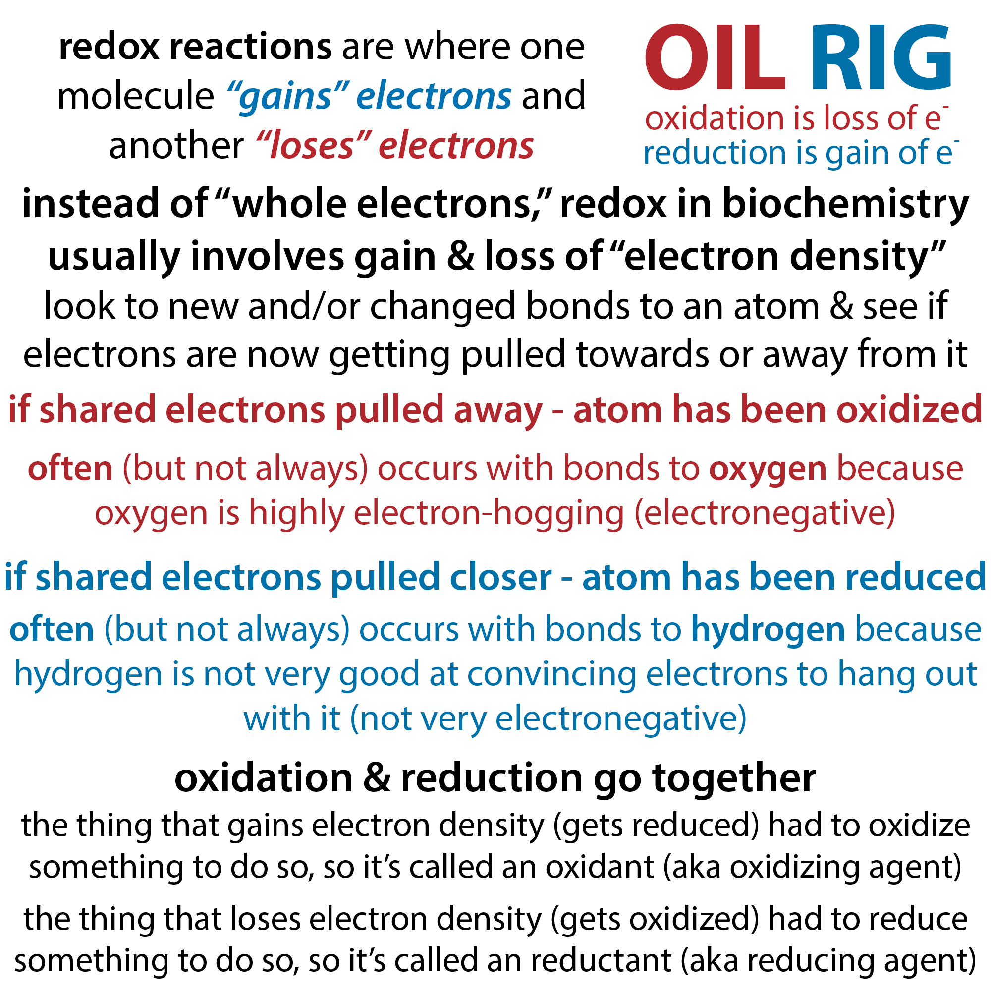
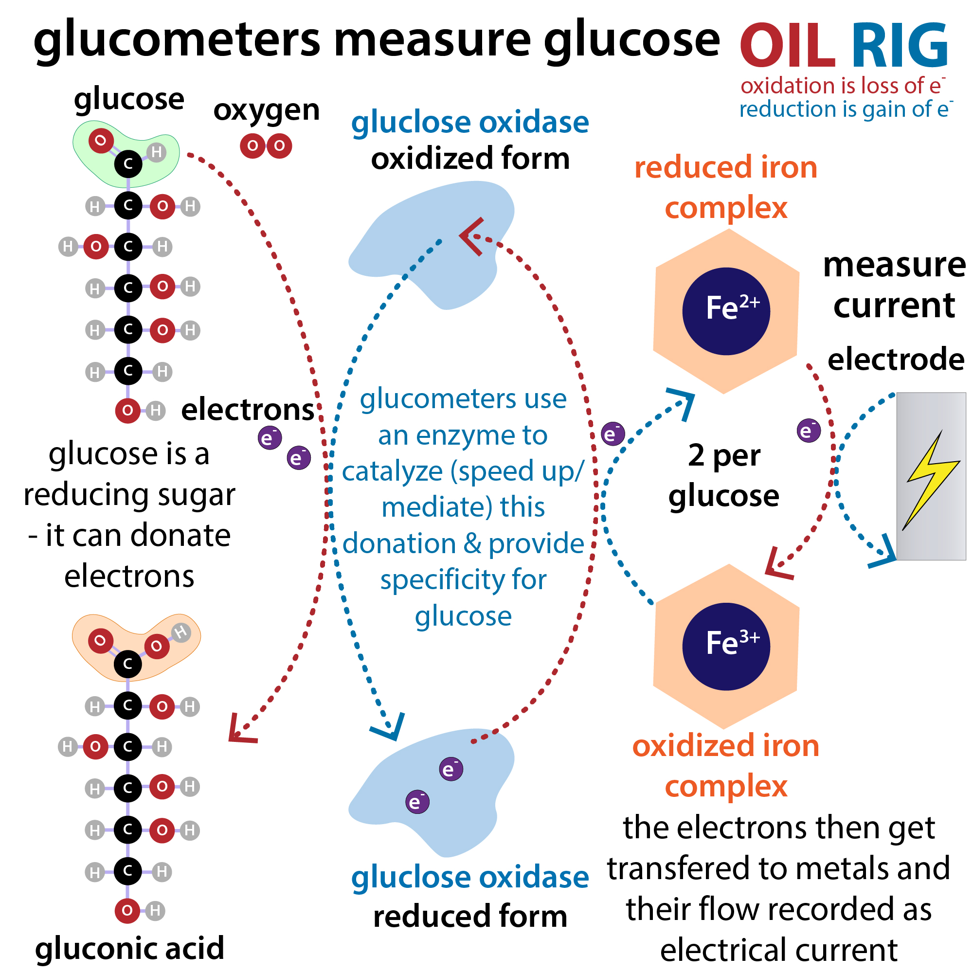
Much more on this in other posts, but basically, molecules interact through their electrons (negatively-charged subatomic particles that whizz around the atoms’ positively-charged nucleus). Reduction and oxidation (redox) are just fancy terms to describe the movement of electrons between them.
You can remember which is which with OIL RIG: Oxidation Is Loss (of electrons) and Reduction is Gain (of electrons). Much more here: blog form – https://bit.ly/redoxbiochem ; YouTube https://youtu.be/reK62_lZJPs
Reducing agents are agents of reduction – they cause something to be reduced and are oxidized in the process. So in order to serve as a reducing agent, a molecule has to be able to be willing and able to give up an electron(s), reducing the thing it gives it to and getting oxidized in the process.
Since glucose is a reducing agent, it will donate electrons, and if we can measure how many electrons are getting transferred – either directly (electron movement is current so you can measure current produced) or indirectly (e.g. transfer them from something that changes color upon losing them) – we can figure out how much glucose there is. The former is the rationale behind the glucose monitors diabetics often use (glucometers) whereas the latter is commonly utilized in organic chemistry labs with the color-changer being a metal in tests like Benedict’s test.
Much more on how glucometers work and why glucose is a “reducing sugar” here: http://bit.ly/glucometers
But here’s the gist. Glucose is definitely not the only reducing agent in your blood so if you want to measure glucose – and only glucose – you can’t just use one of those simple chemical tests. Instead, to get specificity, the tests turn to biological helpers. They use strips coated with an enzyme (glucose oxidase or glucose dehydrogenase) that will take electrons from glucose – and “only” glucose – and pass them off to a “mediator.”
This mediator is often a metal complex like potassium ferricyanide, [K3Fe(CN)6] (aka Prussian red) which can accept an electron, making it ferrocyanide. And then each mediator “ships” the electron to a + charged electrode. Moving electrons is electricity, so you generate an electric current. And this is what gets measured. The more glucose there is, the more electrons can be given up and therefore the greater the current and the greater the signal. Then the computer in the glucometer converts that measurement of current into a measurement of glucose.
Current glucose levels that is. Which is great if you’re trying to figure out whether and how much insulin to inject or sugar to eat. But what if you want to get a sense of blood sugar levels over time? This is where something called “HbA1c” comes in…
Much more on it here: blog: https://bit.ly/hemoglobinA1cbiochemistry ; YouTube: https://youtu.be/sQqWM4iSJ2c
But basically, especially when there’s a lot of it hanging around in the blood, glucose can latch onto the end of a hemoglobin protein chain (hemoglobin, abbreviated Hb, is that protein that carries oxygen through your blood & your red blood cells are chock full of it!) This latching-on of glucose occurs through something called a Schiff base linkage, which rearranges a little to give you a characteristic glycated hemoglobin that can be measured and which is called HbA1c. These measurements are useful because the glycation is “permanent” and red blood cells (where all that hemoglobin & sugar’s at) have a lifetime of a few months. So the A1c levels give you an idea about the blood sugar levels during the last few months as opposed to just “now” and therefore can give doctors information about how well someone’s blood sugar levels are being controlled and whether they might have prediabetes or diabetes or no longer have diabetes (if type 2).
That isn’t helpful for letting you know what to do now, right this moment. But it can tell you how well a long-term treatment strategy is working. So let’s talk about treatment – more specifically just insulin (and disclaimer as always that I’m not a medical doctor, just a geek type of doctor, and if you have medical questions ask a medical doctor).
There are actually multiple forms of insulin that can be used. Some are fast-acting and others are long-lasting. To understand why we need to talk a bit about insulin’s structure.
Much more on this here: http://bit.ly/insulindiabetes
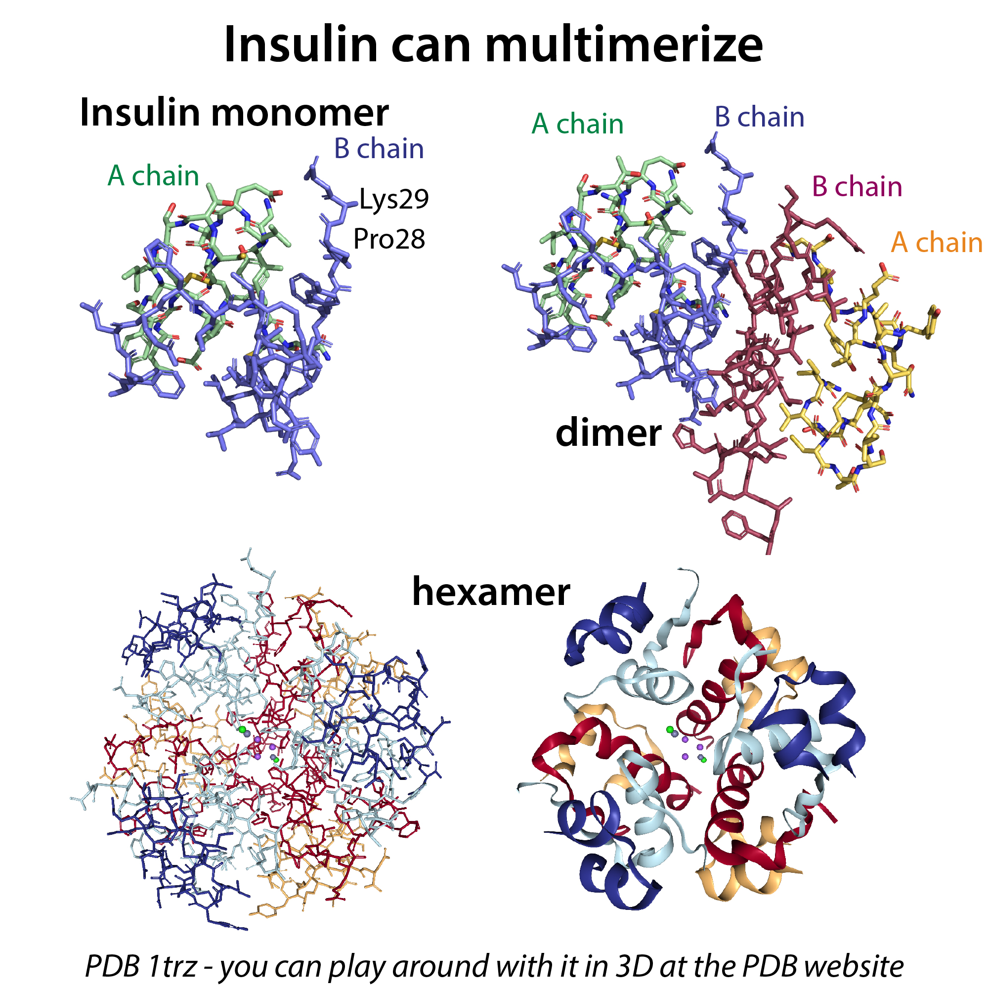
But, basically, one of the main differences between these forms is their tendency to “oligeromize” – individual insulin units (monomers) can pair up to form dimers and then 3 of those dimers can stick together to form hexamers. It’s the monomer form that’s biologically active, so encouraging this form makes for faster-acting insulin, whereas encouraging the hexameter form slows things down.
The reason the hexamers form starts with the reason dimers form, which involves H-bonding between the B chains. And here’s where the nomenclature starts to get a bit tricky. I’ve been using the term “chain” to refer to the polypeptide chains encoded for in a protein’s genetic instructors, which the ribosome faithfully links together. Each protein is synthesized as a chain like this. And usually this chain just folds up into a functional protein. Sometimes grouping up with other chains, but that chain stays that chain.
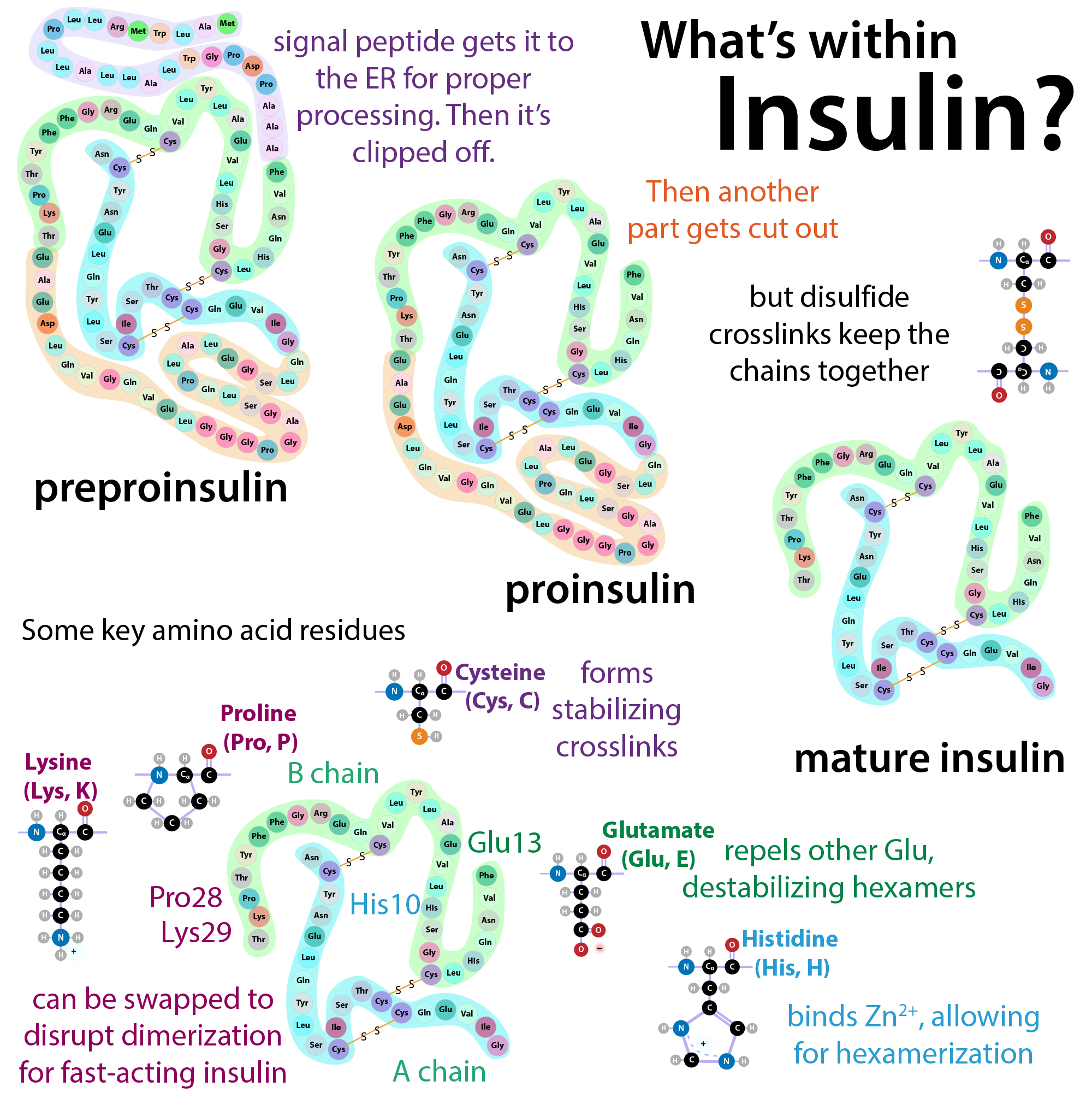
However, in the case of insulin, it gets made as a longer chain called preproinsulin that gets cleaved in 2 places to give you mature insulin which is made up of 2 stuck together smaller chains in each “monomer” of insulin. The first 24 amino acids form a “signal peptide” – As the protein gets made, these come out of the ribosomal tunnel first and signal to the cell that this protein is destined for secretion (getting shipped out). For such secreted proteins, processing usually takes place in a special compartment in the cell called the endoplasmic reticulum (ER). So the ribosome sends the finished chain in there. And then, since it’s no longer needed, the signaling peptide gets cleaved off, leaving you with proinsulin. This pro-insulin then folds up, and gets cleaved again to give you 2 chains, alpha (21 amino acids) & beta (30 amino acids) (both from that original chain).
These chains stay stuck to one another because they have 2 key disulfide crosslinks. Unlike most side chain interactions, disulfide bonds, which can form between cysteine residues (eg. protein-SH + HS-protein -> protein-S-S-protein) are covalent bonds. So they’re strong. And keep the strands stuck together even though their backbone’s broken. So each mature insulin “monomer” is 51 amino acids in 2 chains from 1 original chain. This is *not* the dimer.
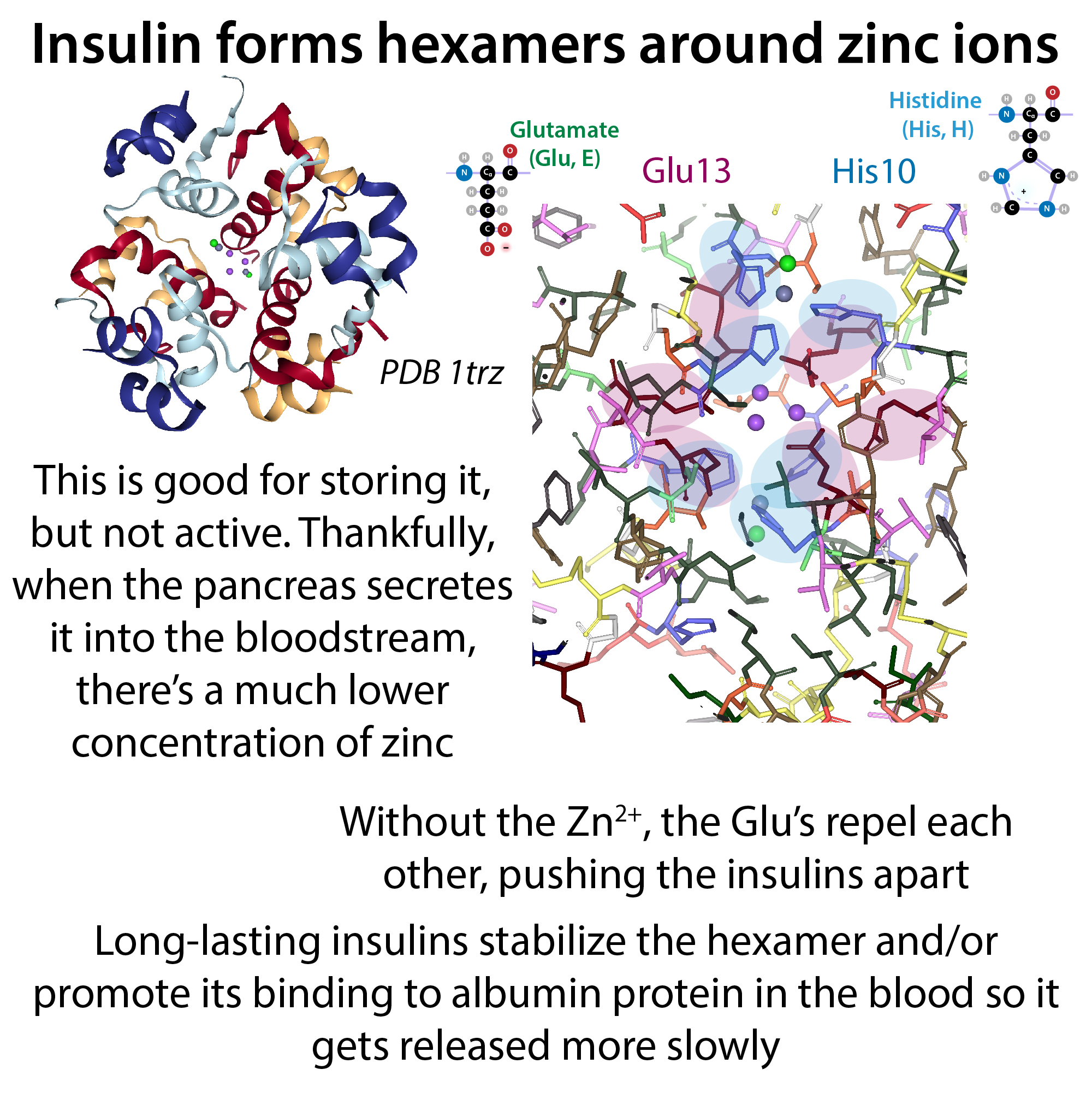
Your pancreas has to stock up on a lot of insulin so it has it ready to ship out when needed. But you don’t want it taking up a bunch of space and you don’t want it “breaking loose” so it makes biological sense to store it in a compact, inactive form. The hexametric form is great for this purpose. When insulin gets secreted from the pancreas into the bloodstream, the hexamers fall apart into the active monomers because the zinc concentration is way lower in the blood.
But turns out that the hexamer-izing can be a problem if you want to use insulin as a drug to treat diabetes. Because, while the monomers and dimers can easily diffuse into the bloodstream, those big ole hexamers have a harder time getting in there if you just inject them under the skin (subcutaneously). To get around this problem, scientists make “designer insulin” thanks to recombinant protein expression technology (being able to stick protein instructions into cells to have them make it for you). Pharma companies can change insulin’s amino acid spelling (primary structure) in a way that doesn’t affect its receptor binding but does prevent dimerization and hexamerization, so that it absorbs better and acts faster. Alternatively, they can change the spelling to promote linking up to make it act slower and last longer so you don’t have to inject it as often.
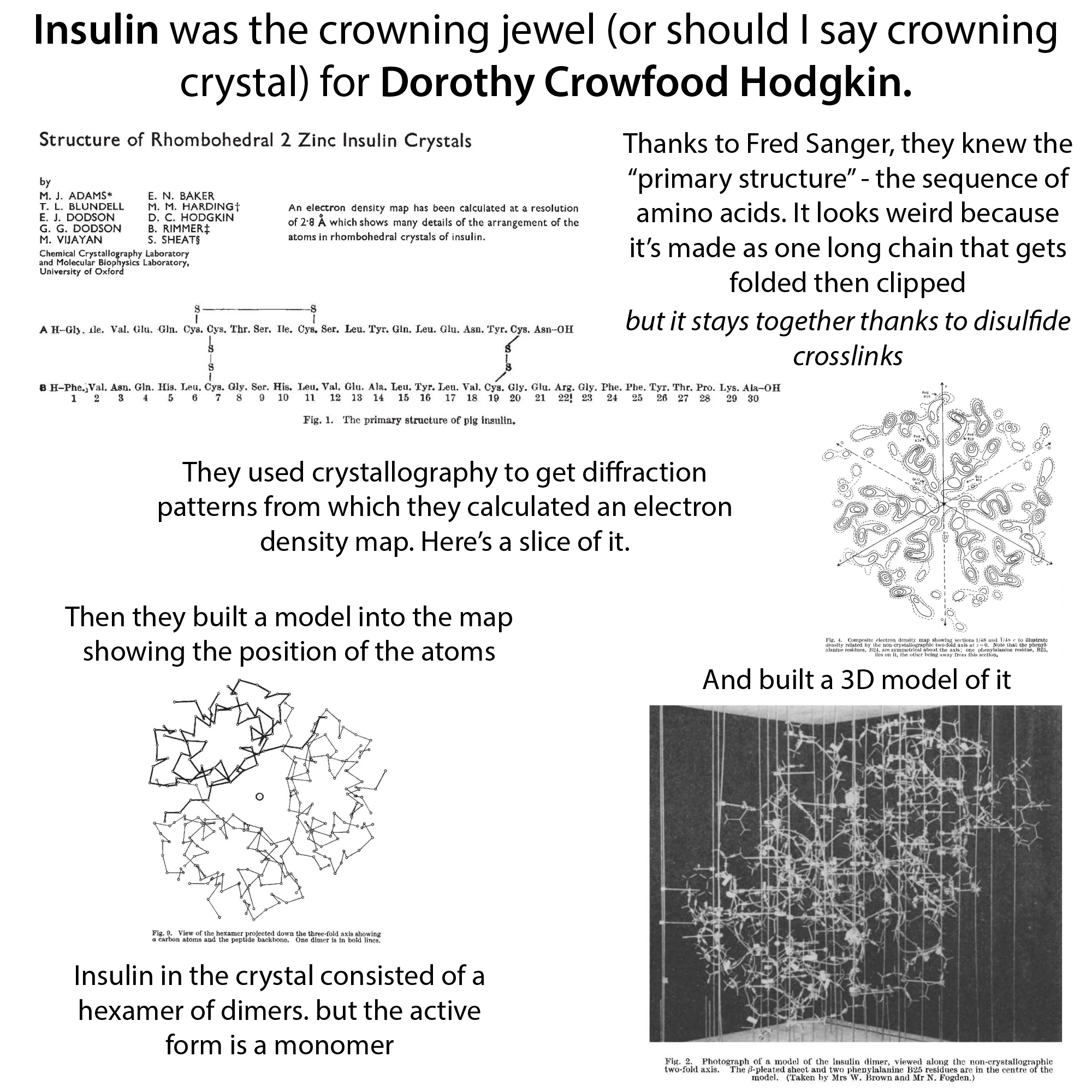
But in order to make those changes, you need to know where to make them, and this is where Dorothy Crowfoot Hodgkin becomes the superhero of our story. Hodgkin was a true pioneer in the technique of x-ray crystallography, which allows you to harness the power of x-rays to figure out what molecules look like at the “atomic scale” (how do all the carbons, etc. link up).
You can learn more about Hodgkin here: http://bit.ly/dorothycrowfoothodgkin
And more about crystallography here: http://bit.ly/xraycrystallography2
Dorothy’s structure showed that the hexamer assembles around 2 zinc atoms in the “2 Zn form”, with the Zn²⁺ held there through “coordination” to a histidine (His, H) from each monomer, His10 (this nomenclature means it’s the 10th amino acid in the chain). Coordinate bonds are a special kind of covalent bond in which one partner (typically a metal) donates a pair of electrons instead of each partner donating a single one (so it’s kinda like the metal is footing the tab for the whole bond, but it’s happy to do so because it has a lot of electrons to spare). One Zn²⁺ coordinates with 3 of the His10’s and the other Zn²⁺ coordinates with the other 3.
Glutamate (Glu, E) 13 in the B chain is at the center of the hexamer and, unlike what you’d expect, the Glu13’s from the different chains are clustered together. Why wouldn’t you expect this? Glutamate is negatively-charged normally. And as we talked about, like charges repel. So you’d think they’d push the insulins apart. But here’s part of where the Zn²⁺ comes in. With its positive charge, it’s able to help neutralize the situation and stabilize the hexamer. So when there’s a lot of Zn²⁺ you get the hexamer form, good for storage (and diffraction) but when Zn levels are lower, like in the bloodstream, the glutamates can repel. Also helping things out, the pH in the blood is higher than in the storage vesicles in the beta cells. Lower pH (greater acidity) means there are fewer H⁺ around to kinda buffer things, so the glutamates really want to get away from each other, therefore helping promote monomerization
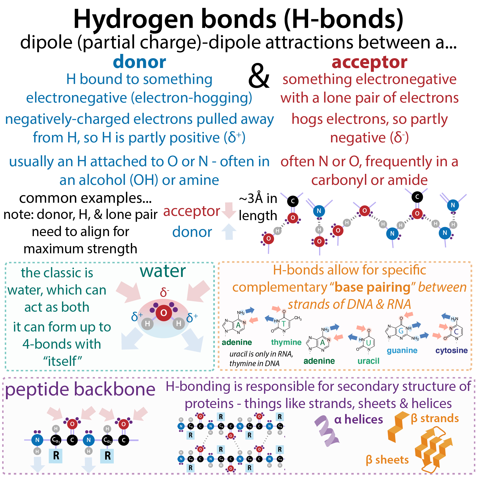
The structure showed that the dimers were forming because of hydrogen-bonding between the C-termini (ending ends) of the B chains of the monomers. Knowing this, once recombinant protein expression was possible, pharma companies could change insulin’s spelling in a way that didn’t affect its receptor binding but did prevent dimerization & hexamerization. For example, insulin lispro (Humalog) swaps 2 C-terminal residues in the B chain, Pro28 & Lys29 to Lys28 & Pro29 (check out PDB entries 1lph or 2kjj). Another one, NovoRapid, mutates a proline to an aspartate. As you might remember from #20DaysOfAminoAcids, Proline (Pro, P) is the least flexible amino acid because its side chain loops in on itself, rebinding to the backbone. So changing this to aspartate (Asp, E) gives greater flexibility and favors monomerization (PDB entries 1zeg, 1zeh or 1kei)
There are also forms to make insulin slower-acting so you don’t have to inject is as frequently. For example, Levemir is insulin covalently bound to a fatty acid – this makes it stick to albumin (a protein that’s at really high concentrations in the blood). The albumin competes with the receptors, leading to longer action. Degludec is a similar version but with stabler hexamers too.
Another cool insulin biochemistry connection – insulin was the first protein to be sequenced. Frederick Sanger and colleagues in the early 1950s figured out the sequence of amino acids in insulin and and developed crucial protein-sequencing techniques along the way. More here: http://bit.ly/insulinsequencing ; YouTube: https://youtu.be/Mi6s0ioOChY