Trypan blue – what’s it do? It gets into dead cells and shows them to you! (It also kills parasites called trypanosomes hence the name…)
Trypan blue has multiple uses but in the lab you’ll find it most often being used in the TC (tissue culture) room, frequently during cell counting (determining the concentrations of cells, usually in order to calculate how much to dilute the cells and/or how much to plate per dish, flask, etc in order to give them enough food (media) and friends (other cells to communicate chemically with to promote growth). We don’t want to count dead cells in our calculations and the Trypan blue dye lets us see which cells are dead so we don’t include them in our count.
Trypan blue is commonly used to test for cell viability (are the cells still alive? What proportion of them?) based on a simple dye exclusion principle. The dye can’t get into live cells with intact membranes (because the negatively-charged membranes repel the negatively-charged dye), but it can get into dead cells or cells with otherwise compromised membranes. Thus, live cells will stay their normal color, but dead cells will turn blue. And when you look at Trypan blue-treated cells under the microscope you can see what proportion of the cells are alive.
This viability determination can have multiple uses. For example, if you see that a lot of cells are dying it can hint at things like:
- Maybe your growth conditions are non-ideal – do you need to supplement the media or something?
- Maybe the chemical you added to test something was toxic to the cells – a further hint is if you do not see death in your control cell (e.g DMSO carrier only)
- Maybe you might have been a little too harsh in your handling of them – remember to pipet gently and stuff
It can also help us only count cells that count when we calculate plating amounts!
When counting cells, we usually want to determine the viable cell concentration. This is often reported in cells/mL or million cells per mL. (1 million cells/mL is the same as 1×10^6 cells/mL)
Traditionally, cell counting has been done manually, using a hemocytomer (a glass slide with a grid) and a little clicker counter to help you keep track). These days, automatic cell counters like the Countess make life a lot easier in a lot of labs. This isn’t a paid endorsement or anything, but it’s what our lab (and an ever-increasing number of labs) use so I will describe it briefly).
The Countess uses little disposable plastic slides with slots for your sample and it does all the counting and viability determination for you. It reports on both the total concentration (including dead cells) and the viable concentration – this is the one you want to use when calculating how much to plate.
No matter whether you’re doing things manually or automated, the sample prep is the same. The dye is typically 2x, meaning you add an equal volume of it to an aliquot (small sample of) your cells. (Usually it’s a 0.4% weight/volume (w/v) % – sorry I wrote w/w in the slide). Typically I add 10 μl cell solution to 10 μl Trypan blue and then pipet 10 μl of that into the Countess. The Countess takes the Trypan dilution into account and adjusts the concentration accordingly so you can use the number it gives you as is, but if you’re doing the counting yourself you need to remember that your sample is actually 2x the concentration you count.
Note: because you’re only counting a super small sample of your cells, it’s crucial that the sample is truly representative of all the cells. So make sure you mix the cells well by pipetting up and down (but gently and without bubbles) before taking a sample and count multiple samples if an accurate concentration is required (a lot of the time ballpark estimates are fine for routine culture maintenance).
Trypan blue is very useful. But it’s not without its flaws. So much so that some people call for it not to be used… (although most of these people are also trying to sell you their alternatives so take it with a grain of salt!)
What’s the problem?
First off, trypan blue is toxic. This is why it can be used as an anti-parasitic. It can wreak havoc if it gets into cells, where it binds to and inhibits lots of proteins. It has a hard time getting into our cells, as we’ve seen, but it gets sucked into trypanosome cells through a process called endocytosis. In endocytosis, cells pinch it a portion of their membrane, transporting things that were on or near the membrane into the cell. Trypanosomes use endocytosis a lot to internalize antibodies that the host’s immune system has sent out to destroy it. Those antibodies recognize surface glycoproteins (sugar-linked proteins) on the trypanosome membrane and bind to them, but then the trypanosome uses endocytosis to swallow the antibodies before the immune system can follow up. In the process, it can swallow a bunch of trypan blue because the dye binds to some receptors on the trypanosome surface.
Pretty neat huh?! I only learned about that today but you can read more here: Alsford, S., Field, M. C., & Horn, D. (2013). Receptor-mediated endocytosis for drug delivery in African trypanosomes: fulfilling Paul Ehrlich’s vision of chemotherapy. Trends in parasitology, 29(5), 207–212. https://doi.org/10.1016/j.pt.2013.03.004
An analog of it called suramin is used to treat trypanosomiasis. Alsford, S., Field, M. C., & Horn, D. (2013). Receptor-mediated endocytosis for drug delivery in African trypanosomes: fulfilling Paul Ehrlich’s vision of chemotherapy. Trends in parasitology, 29(5), 207–212. https://doi.org/10.1016/j.pt.2013.03.004
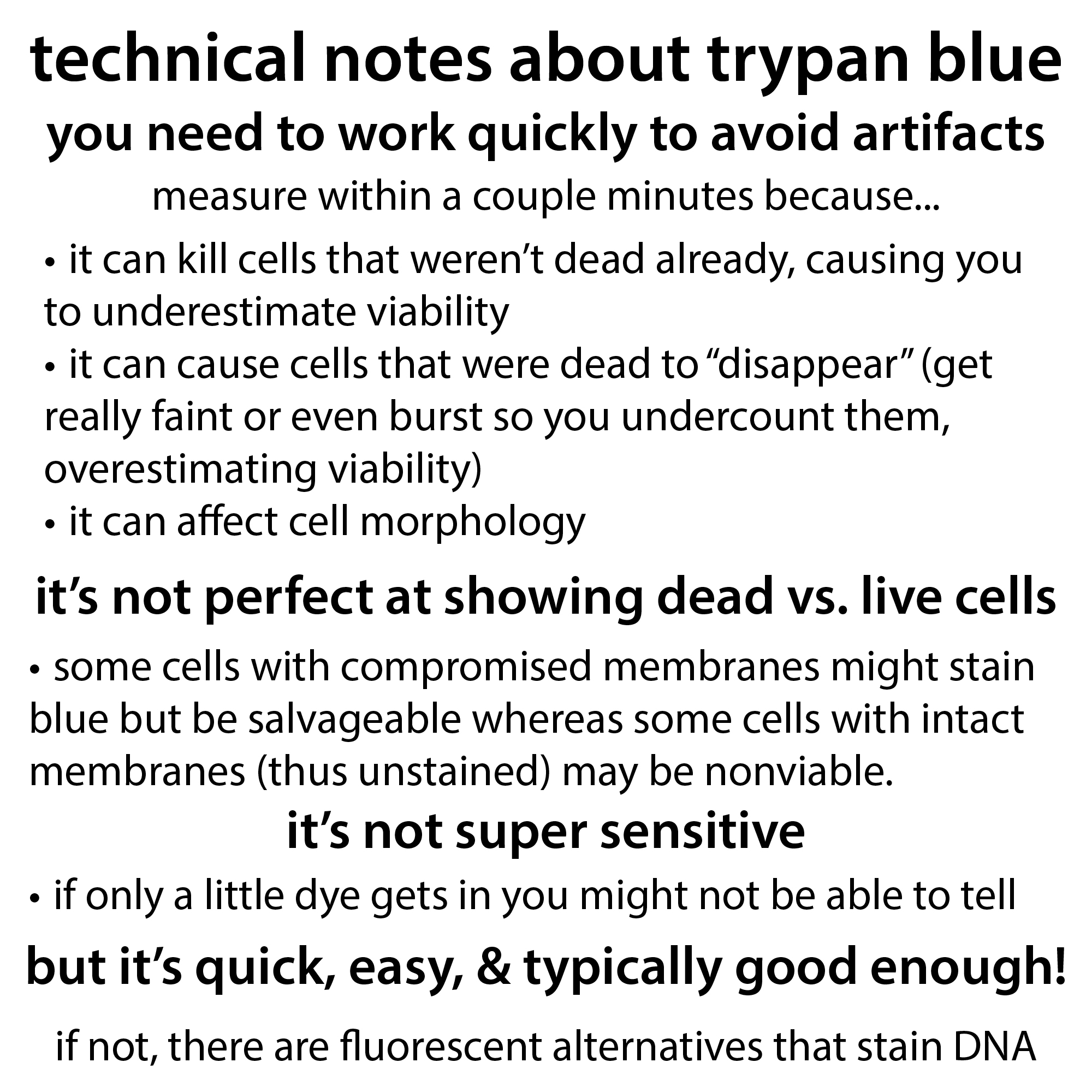
In addition to toxicity issues, trypan blue can cause you to inaccurately count your cells for a few reasons, especially if you wait too long after treating before counting. You want to make sure to count within 5 min of treating. Given long enough the dye can get into even healthy cells, killing them, and therefore causing the measured viability to change over time and causing you to underestimate viability. On the other side of things… Dye that gets into cells can change the cell morphology (shape) and cause it to get diffusely colored – so it turns a really light blue which may lead you to not see it and therefore not count it and therefore overestimate your cell viability.
Chan, L. L., Kuksin, D., Laverty, D. J., Saldi, S., & Qiu, J. (2015). Morphological observation and analysis using automated image cytometry for the comparison of trypan blue and fluorescence-based viability detection method. Cytotechnology, 67(3), 461–473. https://doi.org/10.1007/s10616-014-9704-5
This paper also found the dye could actually cause some dead and dying cells to burst Chan, L. L., Rice, W. L., & Qiu, J. (2020). Observation and quantification of the morphological effect of trypan blue rupturing dead or dying cells. PloS one, 15(1), e0227950. https://doi.org/10.1371/journal.pone.0227950
It’s also important to note that some cells with compromised membranes might be salvageable whereas some cells with intact membranes may be nonviable. So it’s not perfect. It also isn’t super sensitive. A non-viable cell with a mildly compromised membrane might let in just a little dye and you won’t see it.
There are alternatives though. For example, the fluorescent stain propidium iodide, which stains DNA and RNA of damaged cells. And this page also recommends the use of fluorescent stains – a combo of acridine orange (AO) which can get into all cells and stain the DNA and 4′,6-diamidino-2-phenylindole (DAPI) which can only get into dead cells and stains their DNA. But, you then need some way to measure fluorescence, like the device the page is trying to sell you… https://chemometec.com/resources/mini-reviews/why-working-with-trypan-blue-is-not-a-good-idea/
The nice thing about trypan blue is it’s quick, easy, and doesn’t need any fancy equipment to measure it (at least not any fancier than a conventional microscope).
If you’re using more fancy-fancy techniques like flow cytometry, you often use better dyes to assess viability – examples include “dyes that bind to DNA (ethidium monoazide), agents that bind to phosphatidylserine (Annexin V) and amine reactive dyes” Strober W. (2015). Trypan Blue Exclusion Test of Cell Viability. Current protocols in immunology, 111, A3.B.1–A3.B.3. https://doi.org/10.1002/0471142735.ima03bs111
Other techniques determine viability based on the measurement of metabolic activity (for example, are the cells using a lot of ATP? Making a lot of products we can measure?)
Last notes: trypan blue’s full name is 3,3′-[(3,3′-dimethyl(1,1′-biphenyl)-4,4′-diyl)bis(azo)]bis(5-amino-4-hydroxy-2,7-naphthalenedisulfonic acid) tetrasodium salt and, chemically-speaking, it’s an an ionic (negatively-charged) azo dye (it has those N=N groups). It has multiple aromatic rings that allow it to absorb visible light, making it look blue. More on how this all works here: http://bit.ly/lightleafcolor
more about cell culture: https://bit.ly/cell_culture
more about trypsinization: blog: https://bit.ly/trypsin_edta ; YouTube: https://youtu.be/0BgGRKdVOVM
that useful numbers for cell culture guide: Useful numbers for cell culture, ThermoFisher Scientific/Gibco: https://www.thermofisher.com/us/en/home/references/gibco-cell-culture-basics/cell-culture-protocols/cell-culture-useful-numbers.html