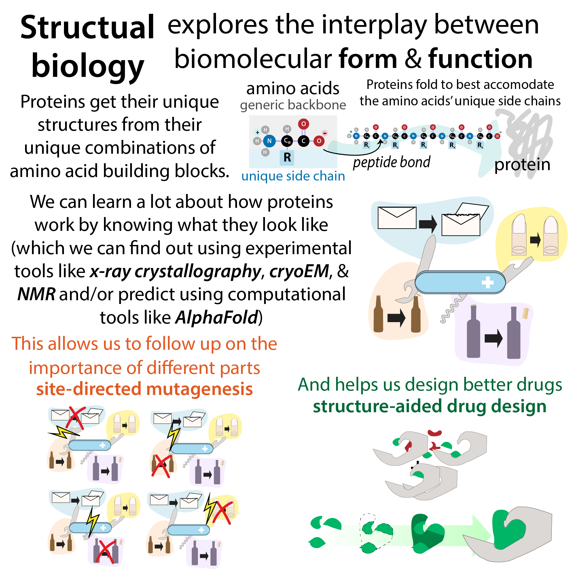
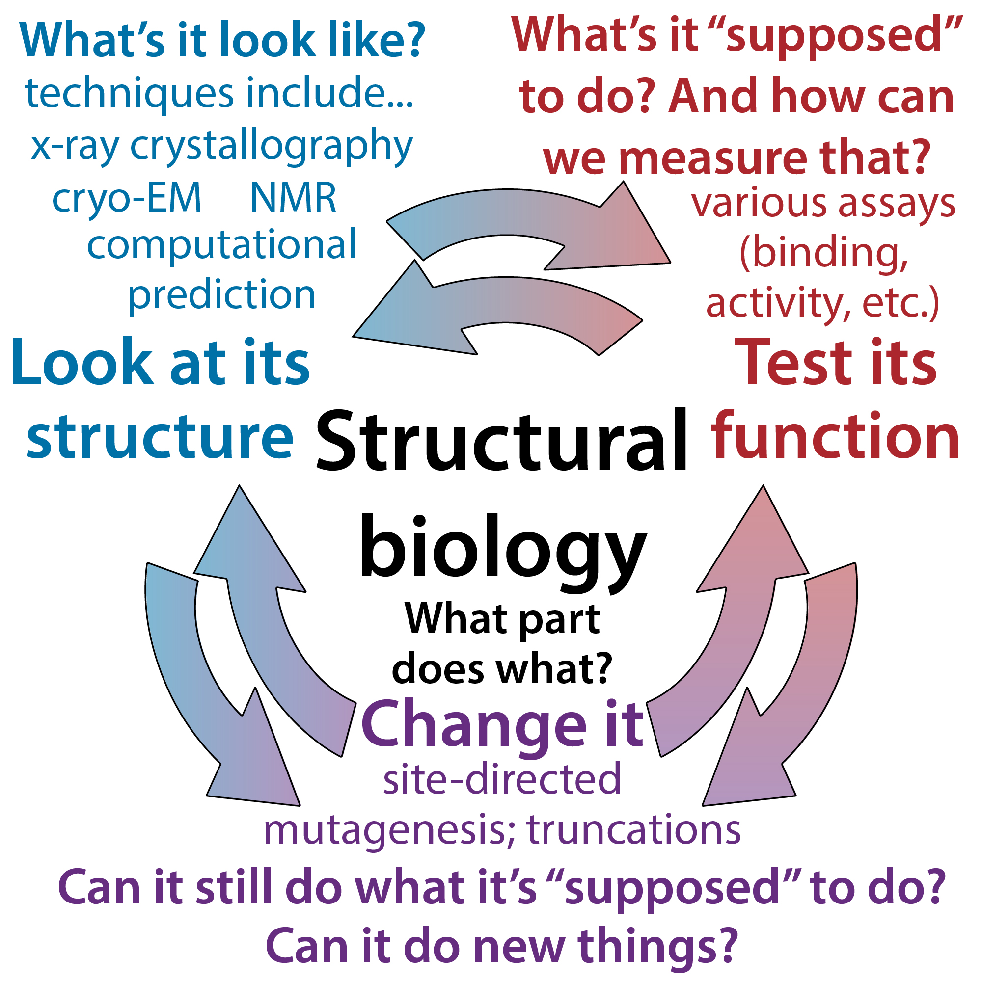
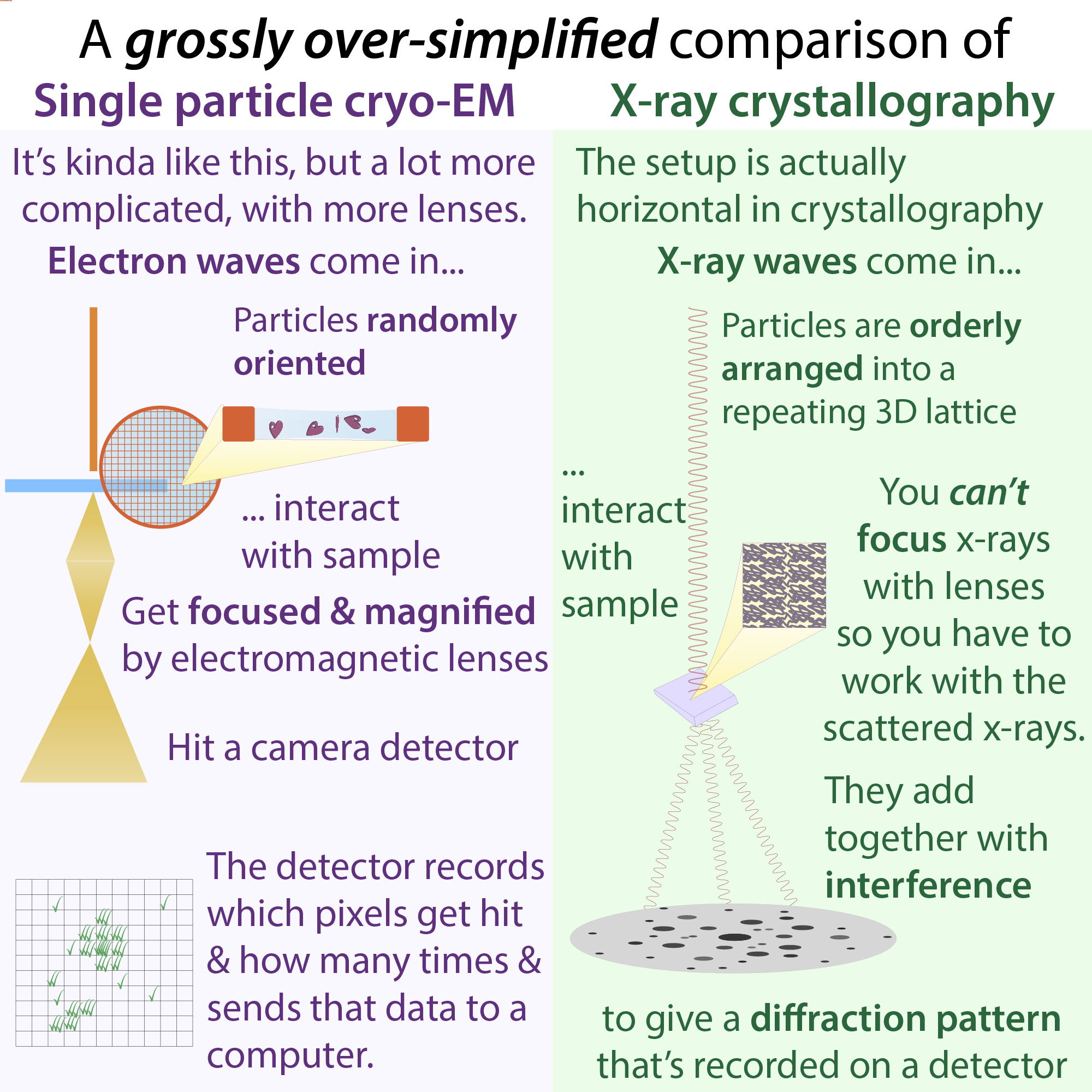
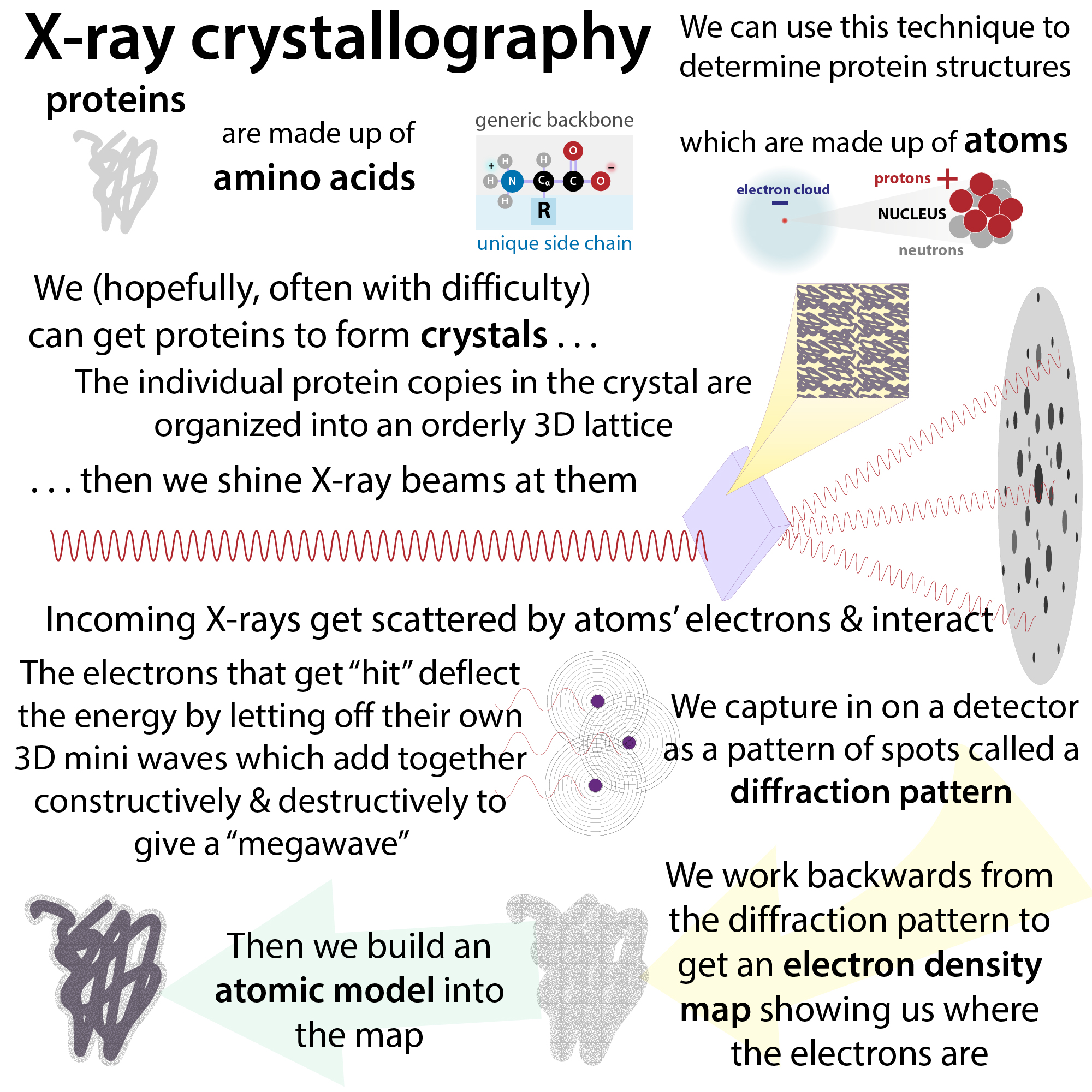
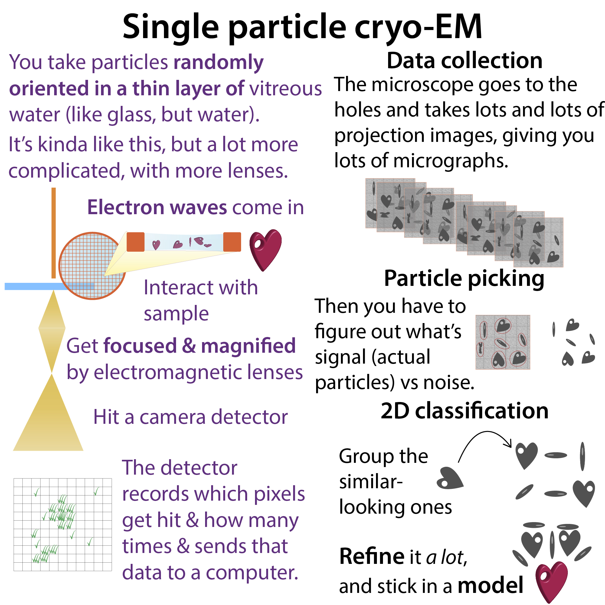
You’ve likely seen a lot of “pictures” of proteins, and it’s structural biologists’ job to take those molecular “pictures” (using methods like x-ray crystallography and cryo-electron microscopy (cryoEM) and study what those “pictures” reveal about how the molecules’ form (structure) relates to the molecules’ functions.
Proteins fold up in a way that makes all of their amino acids happy (e.g. put – charged ones next to + charged ones, let the water-loving hydrophilic ones hang out near the surface and hide the water-avoided hydrophobic ones in the center). Since different proteins have different numbers and combos of amino acids, their amino acid “happy places” will be different, so proteins have unique 3D shapes. And, just like a spoon, a fork, and a knife have shapes that suit their purposes, proteins do too. So having these different shapes enables them to do different things
You can learn a lot about how a protein works by seeing what it looks like (imagine seeing a picture of an open Swiss army knife). And you can also learn about how a protein might “not work” if you can tie up or hide certain parts with another molecule, like a pharmaceutical drug.
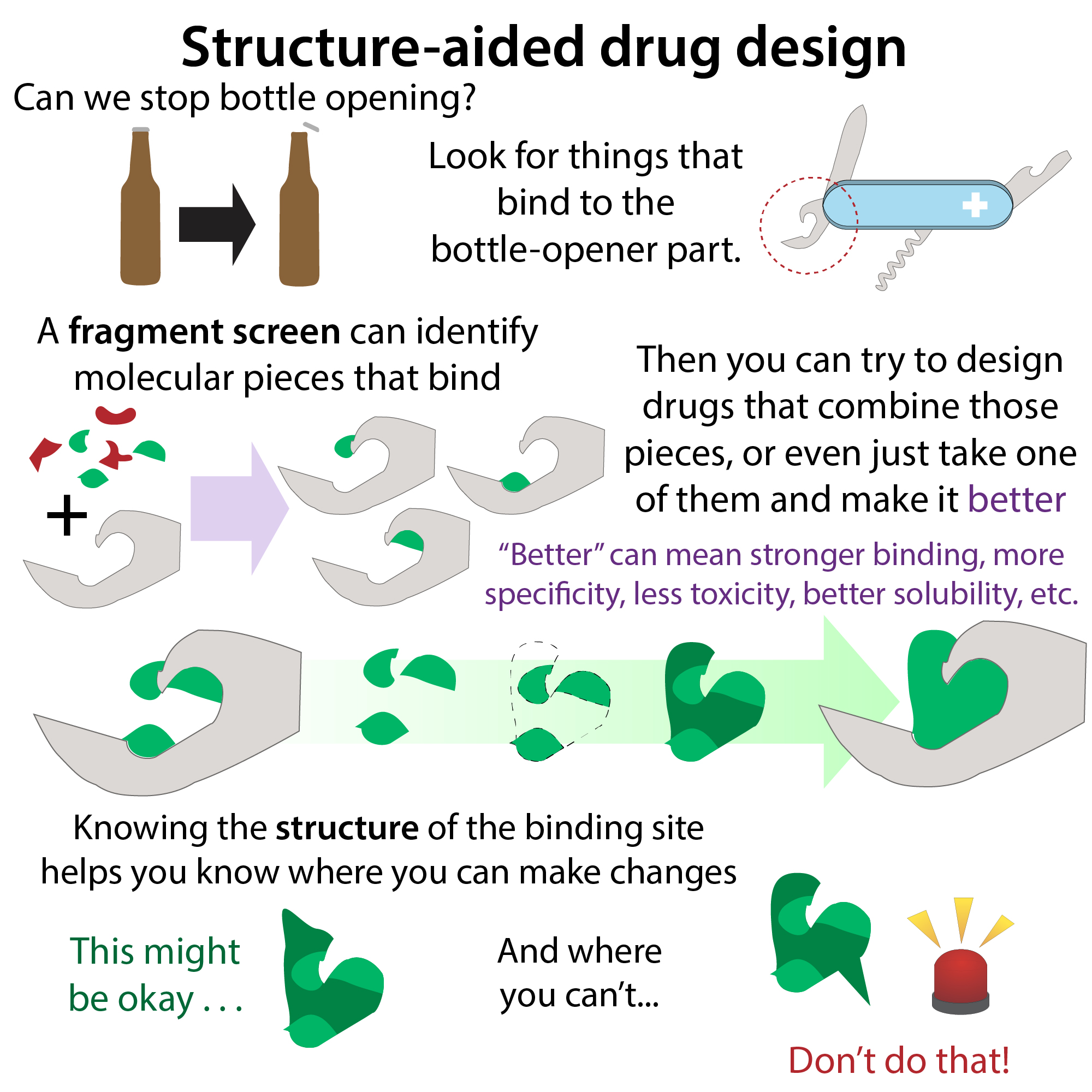
Going back to our Swiss army knife analogy, it’s like if you see a corkscrew and you want to prevent people from de-corking bottles – you could design a “cap” that covers up the pointy tip of the corkscrew. Similarly, if you can see what a protein’s “active site” looks like, the part where the protein “does stuff” (e.g. the place in the protease where it grabs onto and cuts the polypeptide), you can better design a drug that binds there and blocks it.
Instead of designing from scratch, scientists often start by screening pre-existing drugs (some of which are already approved for treating other diseases and thus easier to get approved) and natural products through a compound library screen, or screening pieces (a fragment screen). These screens can be physical screens and/or “in silico” ones that use computer modeling. Such virtual screens, which “dock” compounds or fragments into predicted binding sites, are becoming increasingly common and more sophisticated, incorporating things like molecular dynamics and free energy calculations. These days, scientists can do virtual screens and docking on predicted protein structures, which is wild. . . And must be undertaken with great caution, but shows promise (see referenced review article at end).
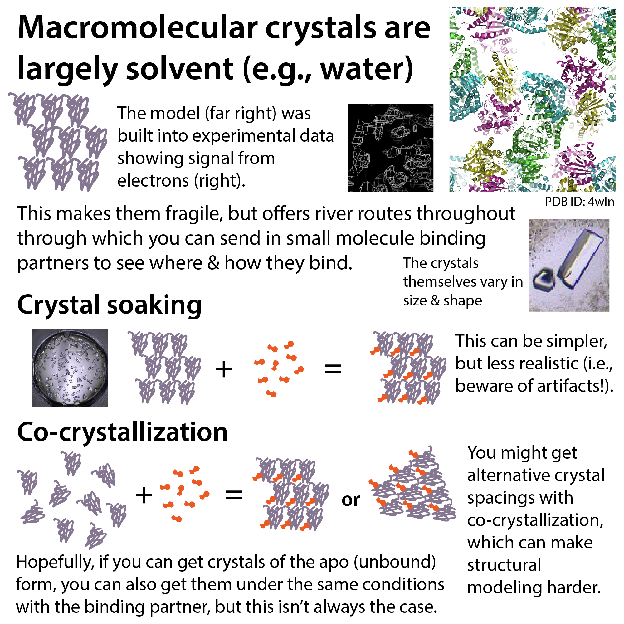
If a protein target is easily crystallizable, scientists might use an XChem fragment screen. There, scientists take huge library of chemical pieces (not full drugs but instead just parts of them), then soak crystals of a target protein in them, then collect x-ray diffraction data from them, work out their structures, and then use the structures of the fragment-bound crystals to try to design therapeutic drugs. Which pieces bind the target site? Which pieces can they combine? How can they modify the bound pieces to make them bind better?
In addition to considering binding optimality, they take into account things like ease of synthesis (it might look great on screen, but you have to be able to actually make it!) and potential toxicity (although you can’t know how toxic something is until you test it, some toxic effects can be predicted based on certain chemical groups the molecules have and similarities to other known drugs).
The top candidates can then be synthesized and then put through additional screens to test for binding activity and strength towards the protein target. Binding’s a prerequisite to having an effect, so it’s a good start. But, if a compound binds well, that doesn’t mean it actually does what they want it to do. So, scientists might then move on to seeing if it can have a desired effect with regard to inhibiting the target activity in vitro, such as with an enzyme assay on purified protein. Then, in cells a dish.
If it passes these “does what they want” tests, it needs to pass the “doesn’t do what you don’t want” test. Off-target activity (effects on other non-intended targets) can cause major toxicity problems. Some of these can be detected in cellular assays, but others don’t emerge until after the drug is metabolized (whereby it might acquire modifications like oxidation that can change its properties) and/or in other cell types, etc. So, scientists might next test if they’re toxic in animals. If they’re not toxic, they can then see if they help animals with the disease. And finally, they can move on to human testing.
Once they have an initial “hit” they can further modify it as needed, adding little pieces here or there to try to give it better solubility, lower toxicity, etc., all the while using that structural information to know which parts they can’t change without messing up the binding-ability.
In addition to using structures of proteins to guide development of small molecule drugs that bind to the proteins, scientists can use structures of proteins to develop better versions of the proteins to use as drugs. Such therapeutic proteins can be referred to as “biologics” or “biosimilars,” which you can learn about here: https://bit.ly/biologics_etc. A
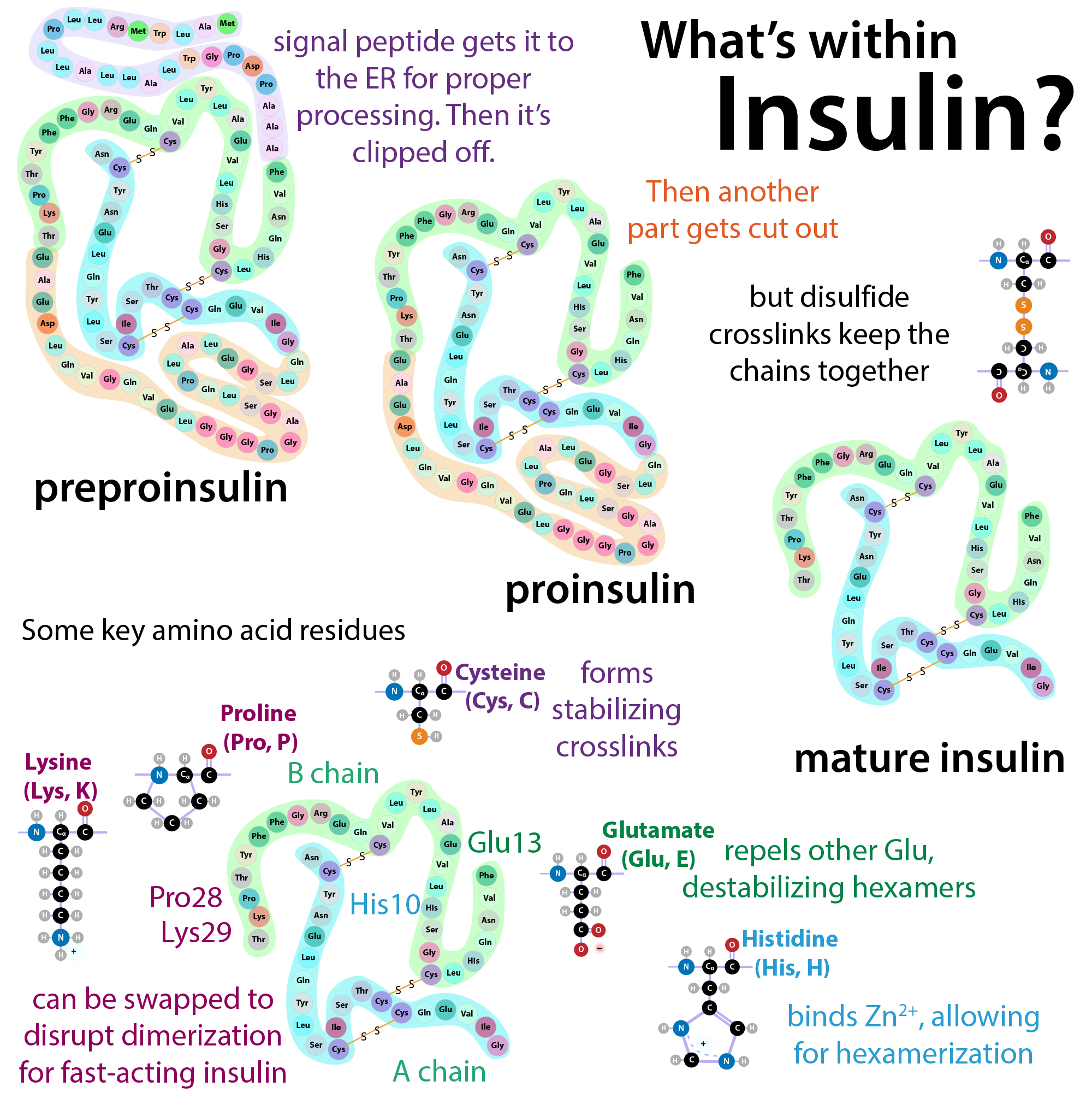
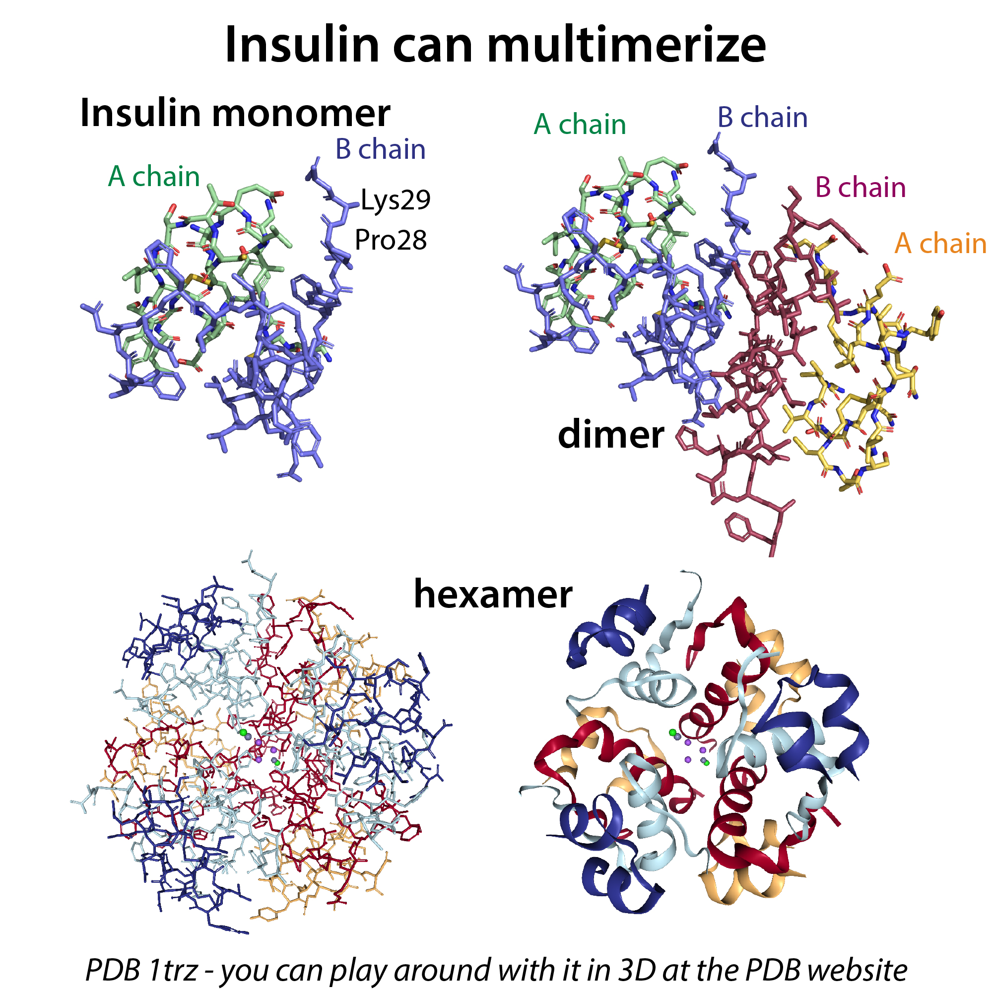
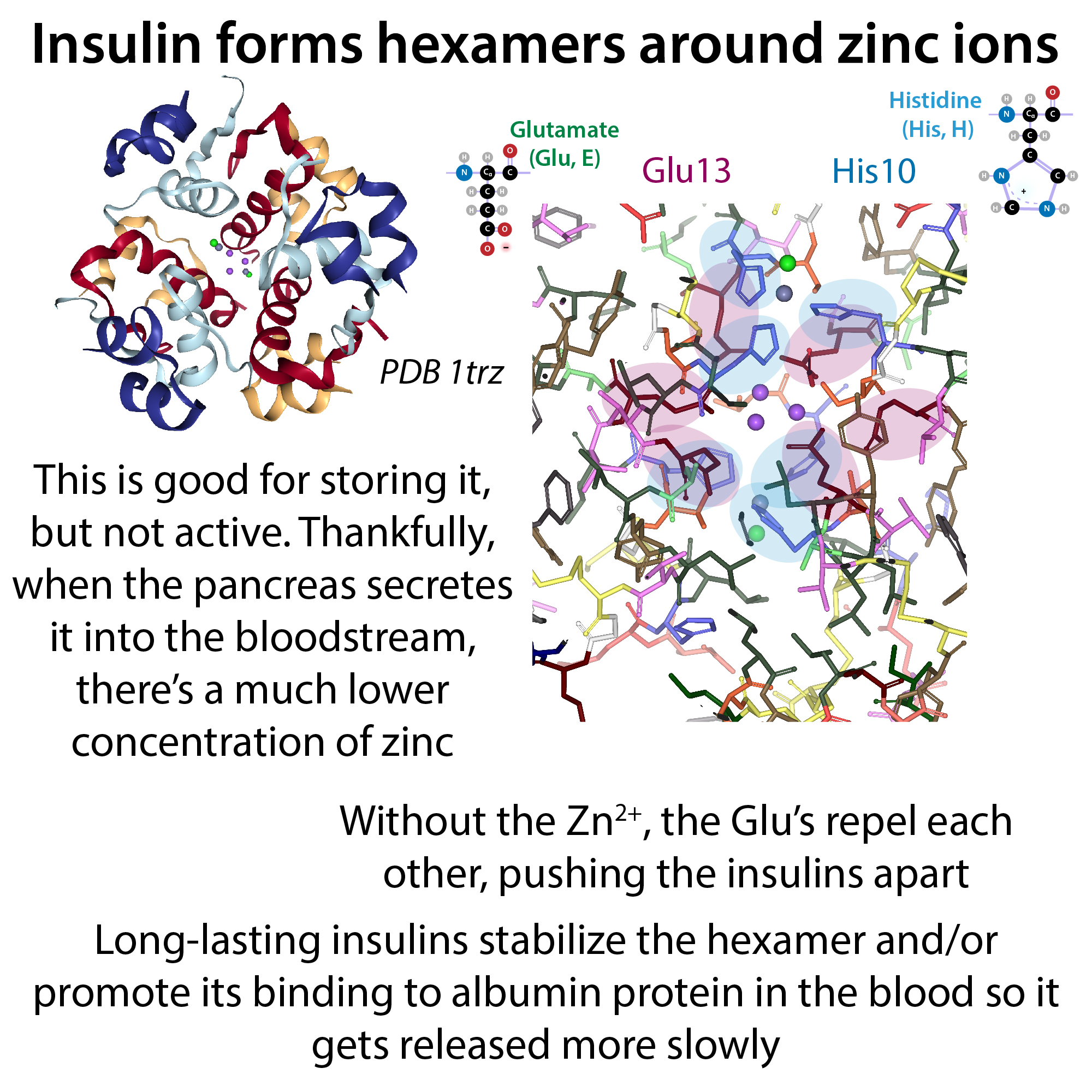
A great example of structure-aided design at work is “designer” insulin. Insulin is a protein that acts as a hormone that helps control blood sugar by telling cells to let in glucose and use it. People with diabetes either don’t make enough of it (Type 1 Diabetes) or their bodies don’t respond enough to it (Type 2 Diabetes). Treatment is therefore often administration of insulins. Scientists use knowledge of insulin’s 3D structure in order to make versions of insulin that are fast-acting and others are long-lasting. Much more on this here: http://bit.ly/insulindiabetes
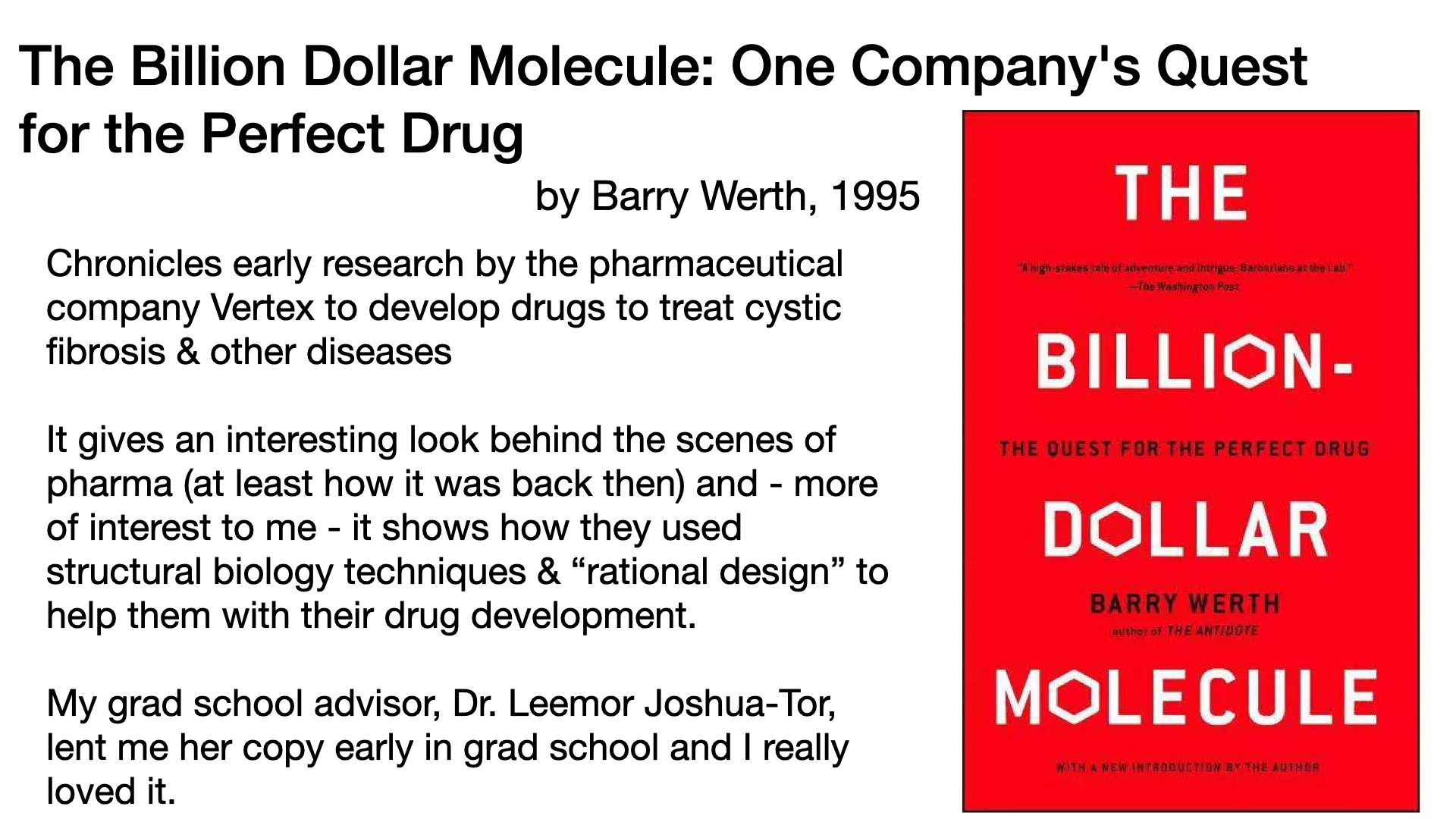
I first was introduced to structure-aided design through a book my PI introduced me to (and lent me) in grad school as part of our structural biology course series: This book really stuck with me when I first read it, and it’s fun to revisit it now that I have years of training: The Billion Dollar Molecule: One Company’s Quest for the Perfect Drug, by Barry Werth, 1995 chronicles early research by the pharmaceutical company Vertex to develop a drug to treat cystic fibrosis. It gives an interesting look behind the scenes of pharma (at least how it was back then) and – more of interest to me – it shows how they used “rational design” with structural biology techniques to help them with their drug development. My grad school advisor, Dr. Leemor Joshua-Tor, lent me her copy early in grad school and I really loved it.
A great account of the history of structure-aided design:
Van Montfort, R. L. M.; Workman, P. Structure-Based Drug Design: Aiming for a Perfect Fit. Essays in Biochemistry 2017, 61 (5), 431–437. https://doi.org/10.1042/EBC20170052
A current look at structure-aided design and the role of computational methods:
Wei, H.; McCammon, J. A. Structure and Dynamics in Drug Discovery. npj Drug Discov. 2024, 1 (1), 1–8. https://doi.org/10.1038/s44386-024-00001-2
And here’s an article describing the use of structure-based design in the development of Paxlovid:
Joyce, R. P.; Hu, V. W.; Wang, J. The History, Mechanism, and Perspectives of Nirmatrelvir (PF-07321332): An Orally Bioavailable Main Protease Inhibitor Used in Combination with Ritonavir to Reduce COVID-19-Related Hospitalizations. Med Chem Res 2022, 31 (10), 1637–1646. https://doi.org/10.1007/s00044-022-02951-6.
A few more:
- Aplin, C.; Milano, S. K.; Zielinski, K. A.; Pollack, L.; Cerione, R. A. Evolving Experimental Techniques for Structure-Based Drug Design. J Phys Chem B 2022, 126 (35), 6599–6607. https://doi.org/10.1021/acs.jpcb.2c04344.
- Van Montfort, R. L. M.; Workman, P. Structure-Based Drug Design: Aiming for a Perfect Fit. Essays in Biochemistry 2017, 61 (5), 431–437. https://doi.org/10.1042/EBC20170052.
- Anderson, A. C. The Process of Structure-Based Drug Design. Chemistry & Biology 2003, 10 (9), 787–797. https://doi.org/10.1016/j.chembiol.2003.09.002.
More posts on structural biology: https://bit.ly/structural_biology
A post on a crowdsourcing effort to develop an MPro inhibitor: https://bit.ly/mproinhibitor