Don’t FRET – here are key things to know about FRET! (Förster or Fluorescent Resonance Energy Transfer) – quick bullet points then the details
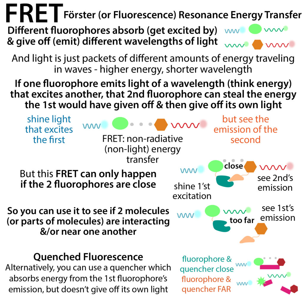
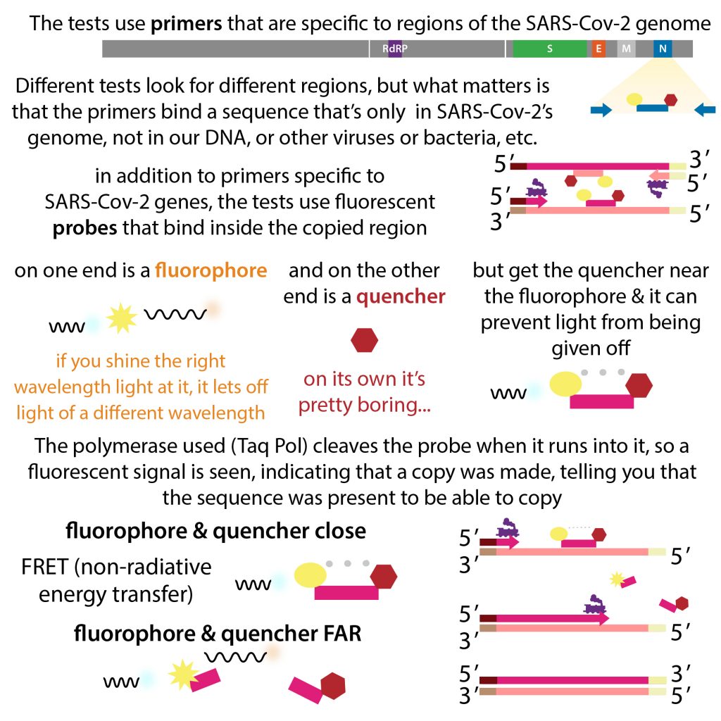
- You have 2 fluorophores – one has an emission wavelength (and thus energy) that matches the other’s absorption wavelength (and thus energy). If they’re really close, and you excite the first one, the energy that would have been given off as light by that first one will instead get stolen by the second one and then the second one will give off light.
- It is distant dependent, with FRET efficiency (E) falling off depending on distance to the sixth power
- You need to be within ~10nm most
- The actual distance depends on a value called R0, which is the 50% transfer efficiency (E) (typically 3-7nm)
- There’s a mathematical equation that relates distance to FRET efficiency for a given pair
- You can use FRET as a “spectroscopic ruler” to measure distances within & between molecules
longer version: http://bit.ly/fretandfluorescence
link to video in case embed isn’t working: https://youtu.be/crA5f1ZGCLo
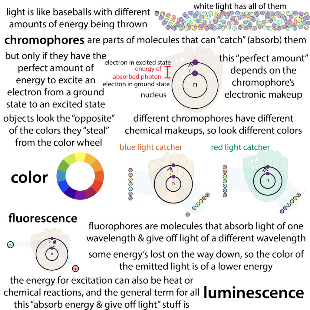
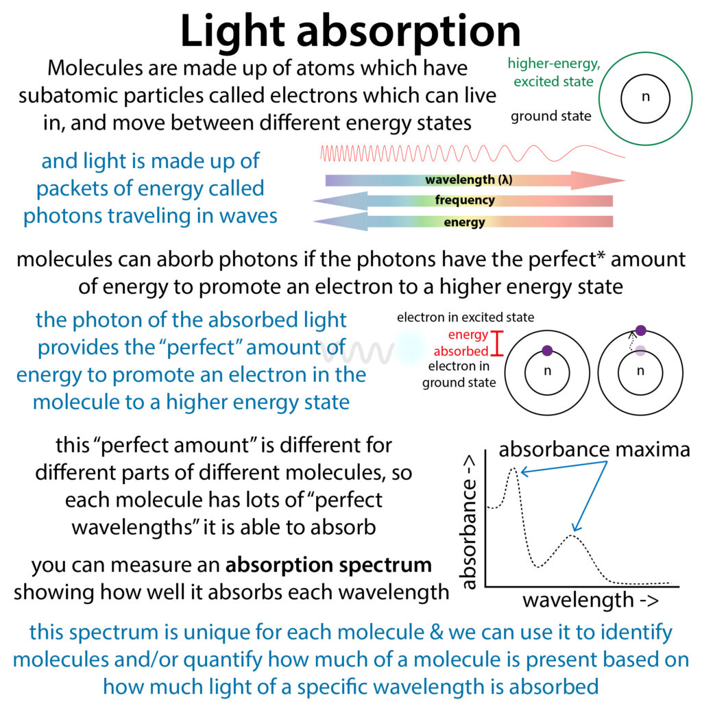
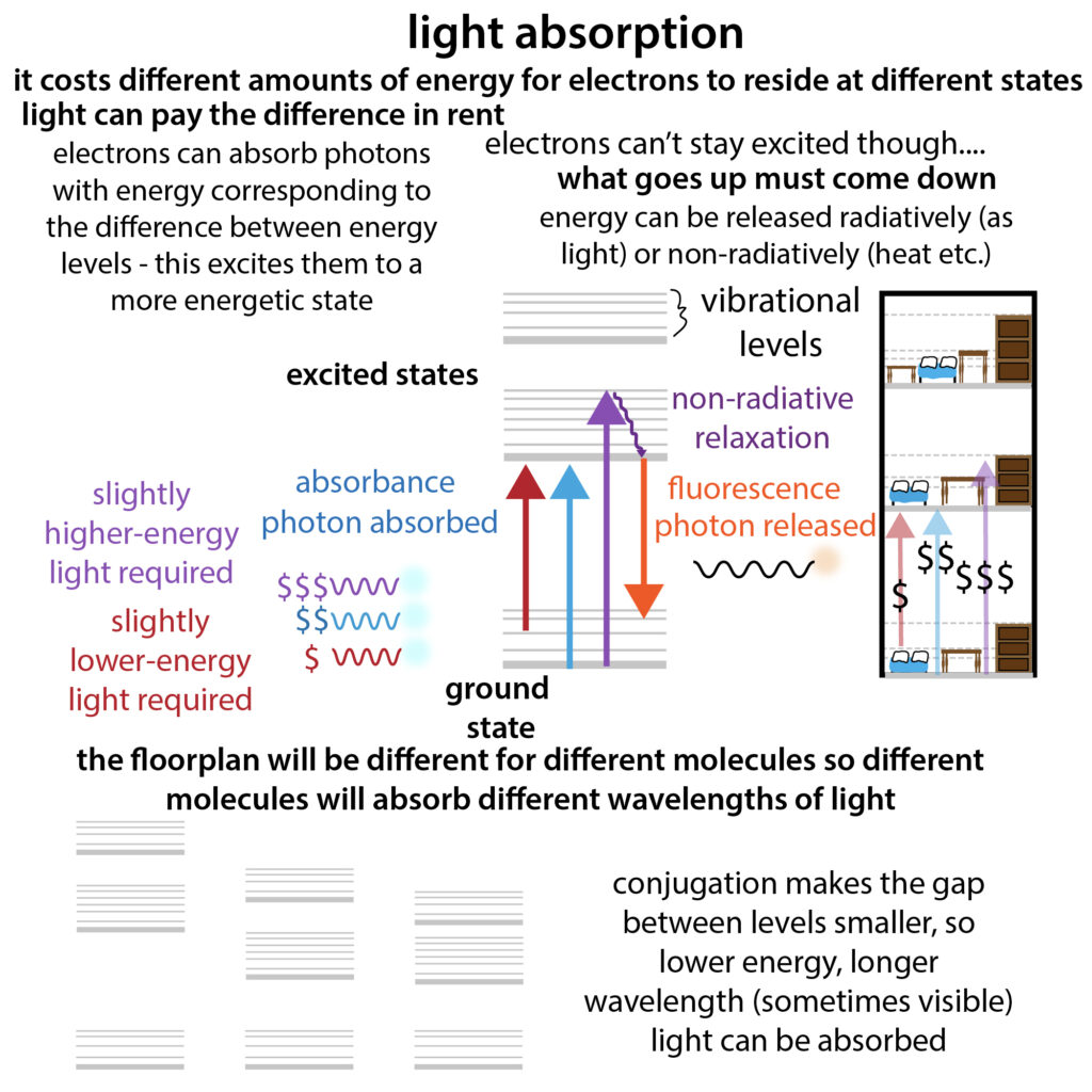
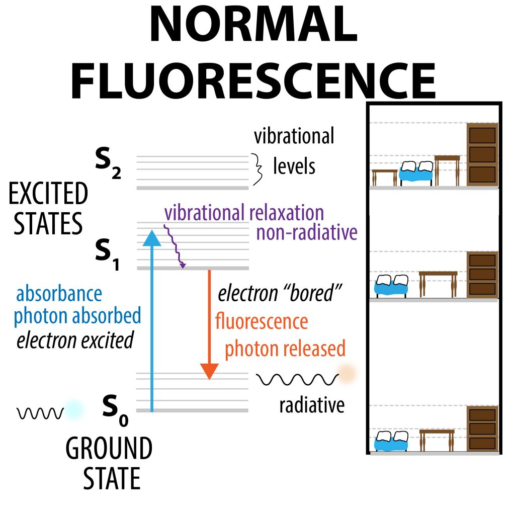
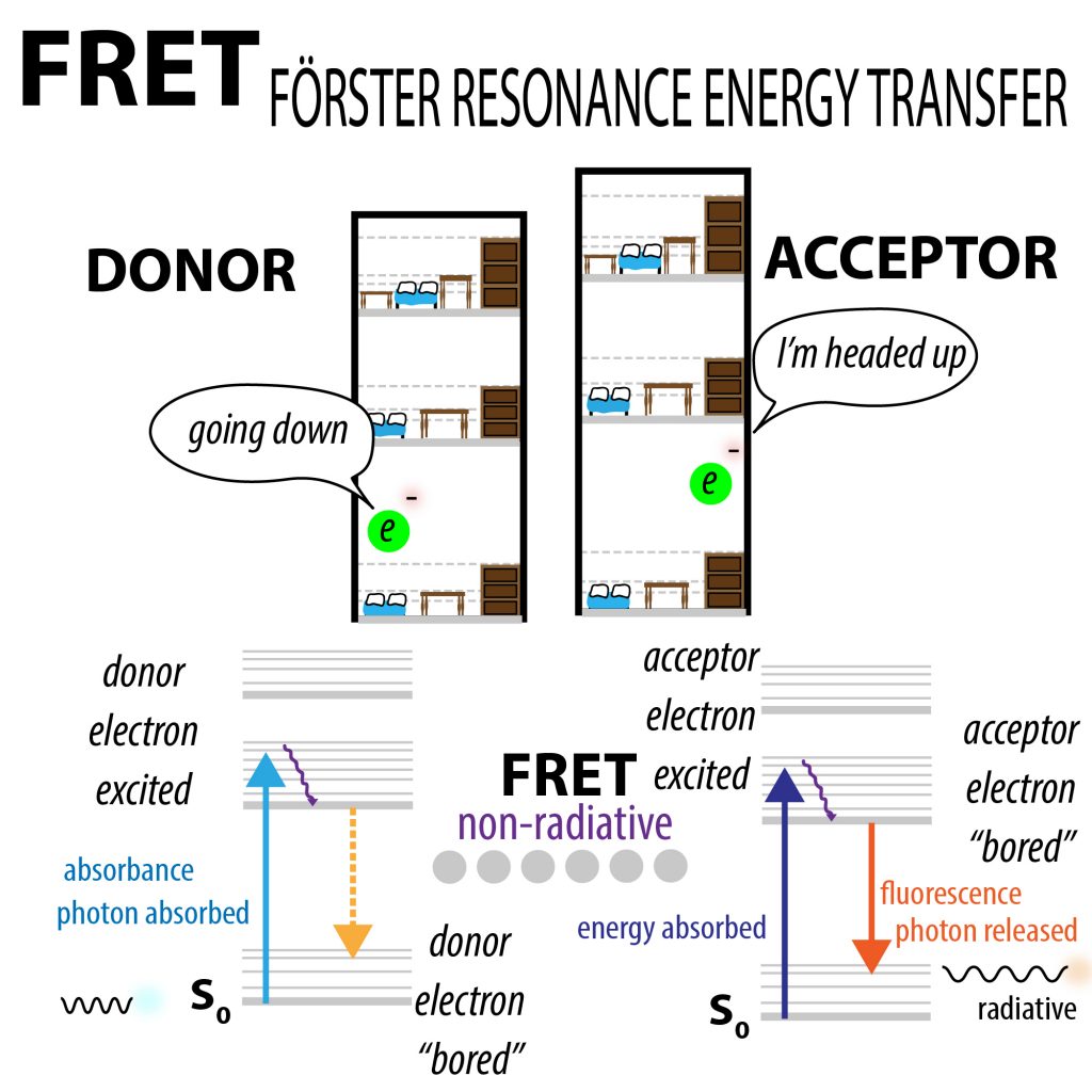
Keep your friends close and your FRET partners closer! The basis of fluorescence is that molecules called fluorophores absorb high-energy light & use some of that energy to excite an electron (like moving it up floors in an electron apartment building where the different floors are different energy states). Then that electron falls back down, releasing lower-energy light that we can detect. In FRET (Förster Resonant Energy Transfer), we couple 2 fluorophores (think 2 side-by-side apartment buildings). Instead of receiving energy from light, the second one receives energy from the first – an electron’s “going down” in apartment building one while an electron’s “going up” in apartment building 2. This can only happen if they’re really close together, so if we see fluorescence from the second fluorophore we can tell that molecules are interacting.
This energy transferring from the 1st to the 2nd isn’t in the form of light (it’s NON-RADIATIVE). It’s transferred through something called Förster Resonance Energy Transfer. There aren’t any particles flowing from one to another (no photons or electrons), just energy “vibes.” If you think of energy as a sort of “money” and photons as “coins,” fluorescence would be like taking in a dime and giving back a nickel (some of the energy gets used for “wiggling” and stuff).
FRET, on the other hand, is more like a sort of wire money transfer, and this sort of transfer can only happen if the molecules are really close. And by close, I mean REALLY close. Visible light has wavelengths of ~380-740 nm (there are 1 billion nm in a m). And FRET can only happen at distances of < 10nm. For some perspective, an “average” human cell is ~ 20,000 nm (20 μm), bond lengths are ~0.15nm and an “average” protein has an ~4-5nm diameter (check out the bionumbers website for some more cool factoids). Note: sometimes, you see values in Angstroms (Å) – an Angstrom is 0.1 nM.
The ability for FRET to occur decreases rapidly with distance – FRET efficiency (E) varies by the inverse sixth power of the distance between them (r). EFRET = 1/[1 + (r/R0)6]
R0 is the Förster radius and it’s the distance at which E is at 50% of its max. This distance is usually a few nm. When r is less than R0, FRET is very efficient, but once you pass R0 things go downhill fast thanks to that “to the sixth” part, with a useful range ~ 0.5-1.5 x R0
This closeness requirement can be really useful. You might have seen microscopy images where people stain cells with a dye that binds to one thing and another dye that binds to another thing and then they overlay the images? It might look like the molecules are really close – they “co-localize”, but they might not be directly interacting. But with FRET, you know they really are.
There are lots of different “versions” of FRET.
Quick note about chromophore and fluorophores. Chromophores are molecules that absorb light. If they give back light, we call them fluorophores. They only absorb and emit light of certain wavelengths, based on their molecular makeup, so each one has a different absorption (excitation) and emission spectrum. More here: http://bit.ly/fretandfluorescence
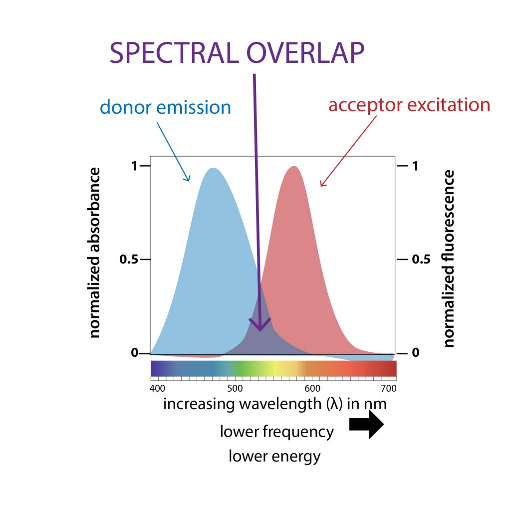
The “classic” is take 2 fluorophores and let one act as a donor to give energy to the acceptor. If you shine a wavelength the donor can absorb the donor can absorb it, and if its emission spectrum overlaps with the acceptor’s absorption spectrum, the acceptor will absorb it.
The acceptor offers an alternative path for giving off energy. So the acceptor, even if it still absorbs the same amount of light as before, will emit less light as before if a FRET partner’s nearby because it’s giving off the energy it absorbs in non-radiative (non-light) transfer to the FRET partner. The donor’s fluorescence is being quenched.
The acceptor on the other hand has a different absorption spectrum than the donor. So it won’t absorb the original light. The “only” way it can fluoresce is if it gets the energy directly from the donor.
So the acceptor won’t emit light in the absence of the donor. Once it gets energy from the donor, it can emit light (we call this sensitized emission) and this light will be at a different wavelength than the light emitted by the donor (but the same as if it absorbed energy directly from a photon)
You can also have acceptors that take in the light but don’t give off light they just release the energy as heat, etc. So you can measure quenching – look at presence or absence of fluorescence versus amounts of fluorescence at 1 wavelength vs the other.
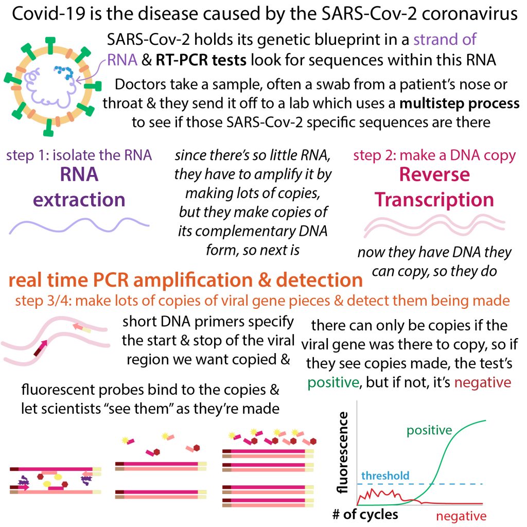
In undergrad, I researched a metallopeptidase (a peptide-cutter). To study its cutting activity I used peptides (short amino acid (protein letter) chains) with a donor fluorophore at one end and an acceptor at the other end. The acceptor wasn’t a fluorophore, so it didn’t give off light of its own – it absorbed energy from the 1st but gave that energy off non-radiatively. We call this sort of molecule a quencher. So if FRET occurred, you couldn’t see anything. And the only way FRET could occur is if the peptide got cut because then the fluorophore was freed from the quencher and could shine.
There are lots of uses of FRET – you can see if molecules interact (e.g. tag 2 proteins with FRET partners), whether molecules change shape (e.g. tag 2 ends of the same protein with FRET partners), etc.
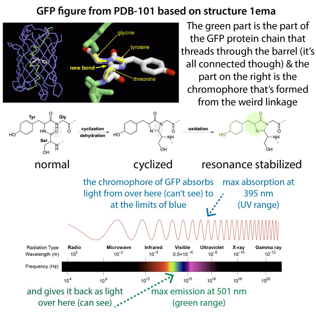
For cell-based assays, you can use molecular cloning to add genes for fluorescent proteins onto the end of the gene for the protein you want to be labeled -> when that gene is made it will have the fluorophore attached. Common fluorescent proteins used for this are optimized versions of CFP and YFP (which are versions of GFP)
For test-tube work you can also use small molecule FRET partners like Cy3 and Cy5, which are frequently used to label DNA or RNA.
more on GFP: https://bit.ly/gfpfunscience
more on FRET & fluorescence: http://bit.ly/fretandfluorescence & http://bit.ly/fluorescentstains
Paper examples:
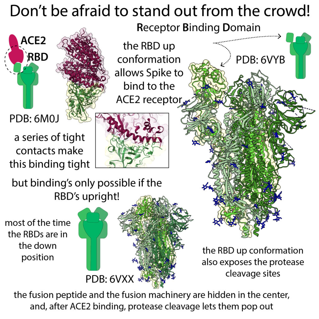
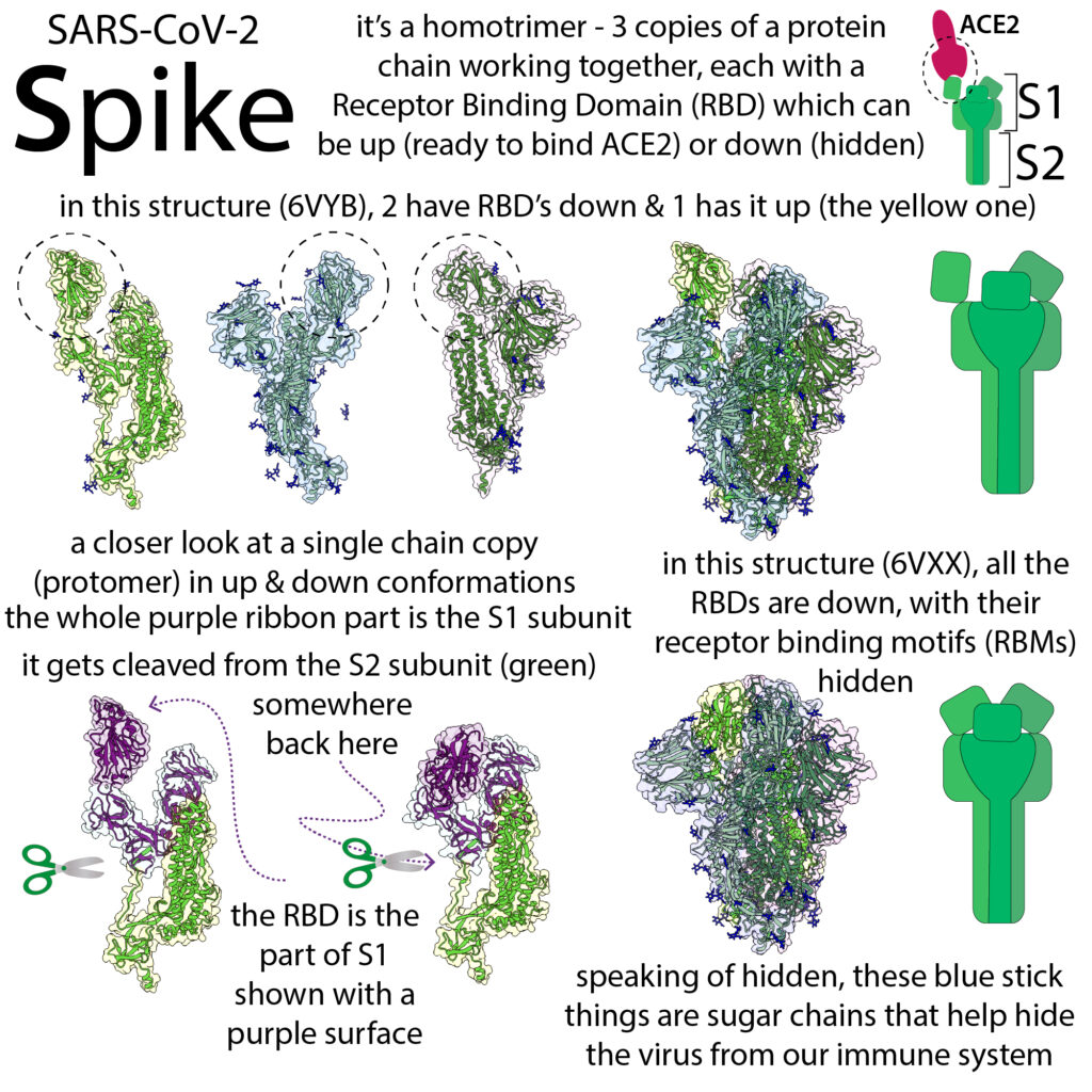
- Cecon, E., Burridge, M., Cao, L., Carter, L., Ravichandran, R., Dam, J., & Jockers, R. (2022). SARS-COV-2 spike binding to ACE2 in living cells monitored by TR-FRET. Cell chemical biology, 29(1), 74–83.e4. https://doi.org/10.1016/j.chembiol.2021.06.008
- Yang, Z., Han, Y., Ding, S., Shi, W., Zhou, T., Finzi, A., Kwong, P. D., Mothes, W., & Lu, M. (2021). SARS-CoV-2 Variants Increase Kinetic Stability of Open Spike Conformations as an Evolutionary Strategy. mBio, 13(1), e0322721. https://doi.org/10.1128/mbio.03227-21
- more on Spike: https://bit.ly/coronavirusspike
FRET resources:
- Bajar, B. T., Wang, E. S., Zhang, S., Lin, M. Z., & Chu, J. (2016). A Guide to Fluorescent Protein FRET Pairs. Sensors (Basel, Switzerland), 16(9), 1488. https://doi.org/10.3390/s16091488
- Benoit Giquel, Addgene, Tips for using FRET in your experiments https://blog.addgene.org/tips-for-using-fret-in-your-experiments
- Cole N. B. (2013). Site-specific protein labeling with SNAP-tags. Current protocols in protein science, 73, 30.1.1–30.1.16. https://doi.org/10.1002/0471140864.ps3001s73
- FRET Basics and Applications an EAMNET teaching module Timo Zimmermann + Stefan Terjung Advanced Light Microscopy Facility European Molecular Biology Laboratory, Heidelberg https://www.med.unc.edu/microscopy/wp-content/uploads/sites/742/2018/06/fret-teaching-module.pdf
- Jares-Erijman, E., Jovin, T. FRET imaging. Nat Biotechnol 21, 1387–1395 (2003). https://doi.org/10.1038/nbt896
- Kim H, Ju J, Lee HN, Chun H, Seong J. Genetically Encoded Biosensors Based on Fluorescent Proteins. Sensors. 2021; 21(3):795. https://doi.org/10.3390/s21030795
- Liu, L., He, F., Yu, Y., & Wang, Y. (2020). Application of FRET Biosensors in Mechanobiology and Mechanopharmacological Screening. Frontiers in bioengineering and biotechnology, 8, 595497. https://doi.org/10.3389/fbioe.2020.595497
- Nikon, Fundamental Principles of Förster Resonance Energy Transfer (FRET) Microscopy with Fluorescent Proteins https://www.microscopyu.com/applications/fret/basics-of-fret-microscopy
- Paul R Selvin (2016), “[illinois] Physics 598 Lecture 5: Still more FRET,” https://nanohub.org/resources/24577
- Roger Tsien Nobel lecture: https://www.nobelprize.org/prizes/chemistry/2008/tsien/lecture/
- Rowland, C. E., Brown, C. W., Medintz, I. L., & Delehanty, J. B. (2015). Intracellular FRET-based probes: a review. Methods and applications in fluorescence, 3(4), 042006. https://doi.org/10.1088/2050-6120/3/4/042006
- Roy, R., Hohng, S. & Ha, T. A practical guide to single-molecule FRET. Nat Methods 5, 507–516 (2008). https://doi.org/10.1038/nmeth.1208
- Verma AK, Noumani A, Yadav AK, Solanki PR. FRET Based Biosensor: Principle Applications Recent Advances and Challenges. Diagnostics. 2023; 13(8):1375. https://doi.org/10.3390/
- Wu, L., , Huang, C., , Emery, B. P., , Sedgwick, A. C., , Bull, S. D., , He, X. P., , Tian, H., , Yoon, J., , Sessler, J. L., , & James, T. D., (2020). Förster resonance energy transfer (FRET)-based small-molecule sensors and imaging agents. Chemical Society reviews, 49(15), 5110–5139. https://doi.org/10.1039/c9cs00318e