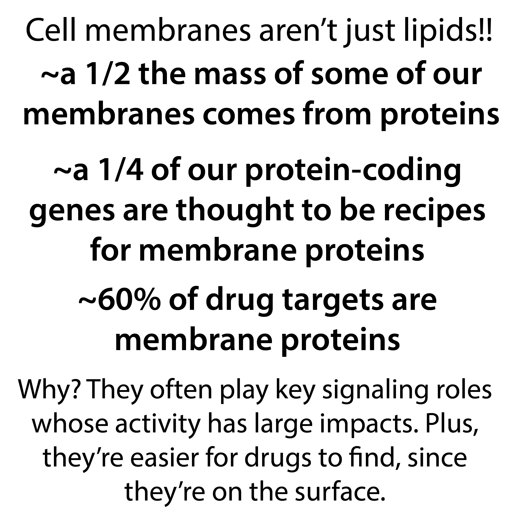
Although we often think about cellular membranes as greasy, oily things, much of the mass of these membranes* comes from proteins – although, since lipids (the fatty parts) are much smaller, there’s 50 times or so more lipid molecules than protein ones.
* mass-wise, protein makes up about 50% for the typical plasma membrane (the membrane separating the cellular interior (cytoplasm) from the extracellular environment, up to 75% or so for some mitochondrial membranes (it takes a lot of protein machinery to generate ATP and this is where it’s done)¹.
YouTube: https://youtu.be/uoXu1EF6atc
These membrane proteins play important roles such as servings as…:
- Receptors – sense what’s going on to help cells respond
- Signal relayers – such as G proteins which are peripheral membrane proteins that pass on messages from integral G protein coupled receptors
- Scaffolds – some membrane proteins help tie together other proteins to create little “factories” where processes can happen that require multiple proteins to find each other and work together
- Transporters – can be active transport (requires energy (e.g. ATP)) or passive (no energy required – just a selective pore or something)
- Cell-cell and cell-surface adhesion molecules – keep cells stuck to one another and/or some substrate like a dish in a lab
- Dockers & fusers – help viral proteins bind to and get into our cells, etc.
Thanks to these functions – and how changes in them can mess things up badly – as well as their easy-to-bump-into location, integral membrane proteins make up ~60% of pharmaceutical drug targets according to some studies².
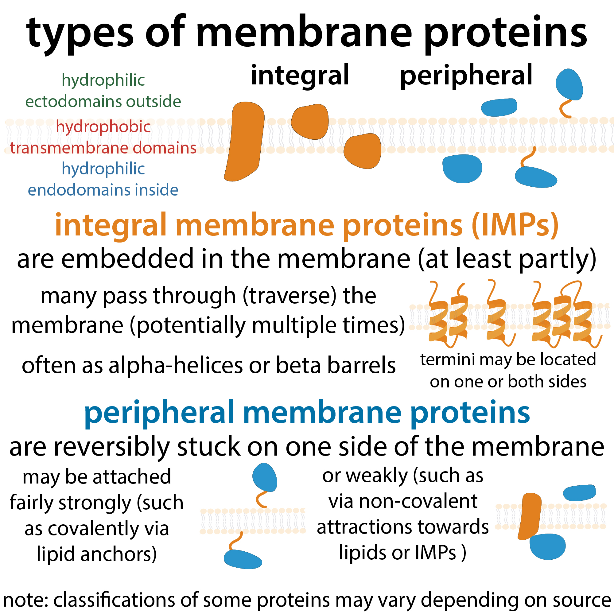
But “membrane proteins” are far from a generic bunch. Instead there’s a bunch of variety! Membrane proteins can be broadly classified into integral membrane proteins, which are “permanently” embedded in a lipid membrane and peripheral membrane proteins which temporarily/reversibly associate with a membrane through interactions with the membrane surface or the surface of integral membrane proteins*. These interactions may be just transient attractions (through electrostatic (charge-based) interactions, hydrogen bonds, etc.) that can be easily disrupted, or they might be stuck more strongly, such as covalently linked to lipids. Those “anchor” linkages can be regulated to strategically release proteins and stuff. It’s pretty cool. But I’m going to focus on the integral membrane proteins in this post. The kind of proteins that you have to really disrupt the membrane (such as with detergents as we’ll see) to “free.”
*warning: the distinction is kinda blurry when it comes to anchored and slightly-embedded proteins – some places classify some things one way that another place classifies another way
Within the ream of integral membrane proteins (IMPs), we can further classify things based on whether or not the protein passes (traverses) all the way through (is a transmembrane protein) or only partway through (is a monotopic IMP). Then, within the transmembrane class, we can classify things based on how many times they traverse the membrane (number of passes) and where the protein ends (the starting, N terminus, and the ending C terminus) are located. If it passes through an odd number of times, it will have on end inside and one end outside (but some have C in, some have N in). With an even number, the ends are both on the same side.
More of the structural details later on since they get kinda in-the-weeds and I don’t want to lose people already. Instead I will start by talking about something more of you can relate to.
Many membrane proteins are glycoproteins – they have sugar chain(s) attached to them. These play some important roles in adhesion, cell signaling, immune recognition or evasion, etc. In an example you may be all too familiar with, viral fusion proteins (IMPs that dock onto cells and allow the virus to fuse with the cellular membrane) such as coronavirus Spike proteins, often are glycosylated which can hide the protein surface from immune recognition.
Speaking of Spike, purification of the Spike protein for research and protein-based vaccine production provides a good opportunity to discuss some of the challenges and strategies for studying integral membrane proteins. Peripheral ones are easier since you can release them from the membrane and they’ll be happy in an aqueous (water-based) environment. But, even if you were to be able to just release an IMP, it would act impish… it wouldn’t be happy in water. Because the transmembrane parts are “designed” to be surrounded by lipids. So you need to keep them surrounded by lipids.
In order to do this, you can use detergents (artificial soaps) to disrupt the membranes and extract the proteins into micelles (little bubble-like things). Let me step back a minute and discuss what membranes are actually made up of so that you can better understand what’s going on…
I know I’ve been focused on how important membrane proteins are, but the lipids are crucial too. Not only do they allow our bodies and our cells to compartmentalize, but they also themselves have different properties and modifications that allow them to interact with proteins and other molecules, etc. Different membranes (e.g. plasma membrane vs. nuclear membrane vs. mitochondrial membrane vs. endoplasmic reticulum membrane) have different lipid compositions. They are all made up of phospholipids, but they have different proportions of different ones.
In a bit more detail, phospholipids are what we call “amphiphilic” – they have parts that water likes (hydrophilic parts) and parts that water hates (hydrophobic parts). The hydrophilic parts of phospholipids are phosphate-containing “heads” (which is where a lot of the variation comes from) and the hydrophobic parts are hydrocarbon “tails” (long chains of carbon & hydrogen). The phospholipids in our membranes have 2 tails, so they have a sort of rectangular shape.
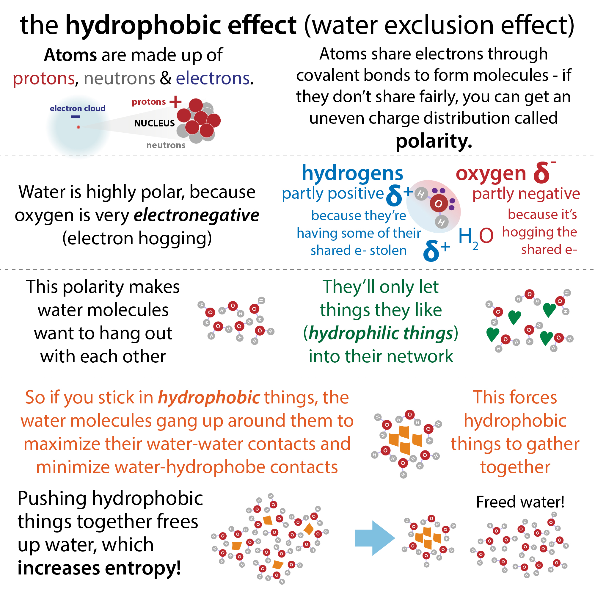
If you stick them in water, the water molecules are going to gang up on them, flocking to the hydrophilic parts (as well as clinging to other water molecules) and running away from the hydrophobic parts. This leaves those hydrophobic tails left with nothing to hang out with but one another. But their rectangular shape makes this awkward. The solution? Take inspiration from a sandwich! The phospholipids form a bilayer membrane where the heads take on the role of the bread and the tails the peanut butter.
Speaking of butter, sorry I didn’t explain more before but the term “lipid” is basically a broad category that encompasses fats, oils, waxes, and other largely “nonpolar” hydrophobic molecules.
Also speaking of butter, the membrane is fluid. Individual lipids can move around within the layer (leaflet) they’re in – but they need the help of proteins called flipases in order to swap to the other leaflet. This allows there to be distinct asymmetry in membranes with the two layers being different. Proteins can also move around in the membrane (though not quite so easily as the lipids), leading to a concept called the “fluid mosaic model” where you have a sort of sea of lipids that proteins live & move around in. And often the proteins hang out in groups – this potential for localization, using the membrane as an organizing center is one of the virtues of membranes.
When we think about membrane proteins there’s a tendency to focus on “main membrane” – the plasma membrane. I know I have this bias myself. But don’t forget those other ones. And don’t forget that the lipid composition (what proportion of the lipids have which head, etc.) can matter greatly. That being said, when we study membrane proteins in vitro (in a test tube or other artificial environment) we typically make simplifications that make things more generic and less realistic. Such as by using those detergents.
So, what is a detergent? It’s an artificial soap. And what’s that? Basically, it’s another type of amphiphilic molecule a lot like a phospholipid, but with a single tail. This makes it more conical than rectangular so it’s less awkward to arrange themselves in a single, spherical, layer. So this is what they do when you stick them in water. They form those liquid-filled bubble things called micelles, where their hydrophilic heads are on the outside, facing the water, and their hydrophobic tails are in the center (this is also where hydrophobic gunk hangs out when you’re using soap or detergent to wash things).
Since detergents “look” like phospholipids and have a lot of the same properties, they can elbow their way into phospholipid membranes, disrupting them and picking up some proteins which they then drag into their micelles. And this offers you a way to extract membrane proteins from membranes. They’re still hard to purify and keep happy though, so scientists often try to avoid all this. Instead, if they want to study membrane proteins outside of a cellular context they often express (have cells make) and purify just the hydrophilic inside (endodomain) or outside (ectodomain) parts.
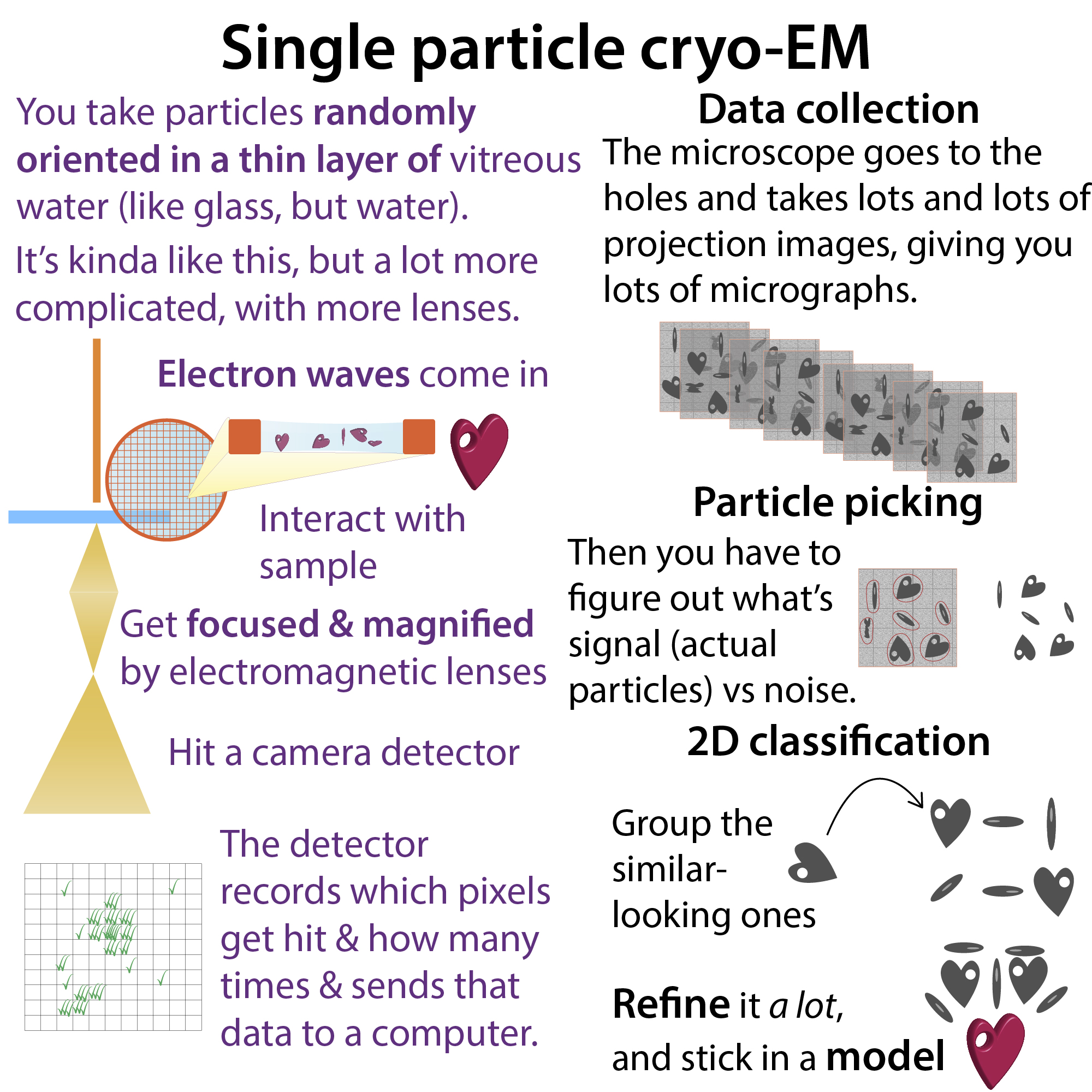
That can be good enough to study things like binding affinity, but you lose a lot of information. A somewhat more realistic environment comes from using lipid nano discs, which are kinda like bilayer lifesavers surrounding proteins. These are frequently used for the structural study of individual membrane proteins or complexes (e.g. via single molecule cryo-electron microscopy (cryoEM)).
note: I have huge respect for people who study membrane proteins. I worked with them during a lab rotation in grad school and man it’s hard! never got those nanodiscs working… but anyways…
For big complexes, structural biologists (scientists interested in seeing what proteins look like at the molecular level) can use a technique called cryo-tomography to look at the membrane as-is, rather than looking at purified complexes. It’s kinda like giving membranes a CT scan and it’s gaining popularity (and “goodness”).
Since we’re on the topic of structure, here’s more on the structural of transmembrane proteins, as promised:
When proteins traverse a membrane, they tend to do it in specific “shapes” – alpha helices or sometimes beta barrels. Both of these terms refer to secondary structural motifs (characteristic shapes regions of proteins adopt due to hydrogen-bonding interactions between atoms in the protein’s backbone). More on these in other posts. For now just think of a spring (alpha helix) and an accordion (beta strands) rolled up into a cylinder (beta barrel).
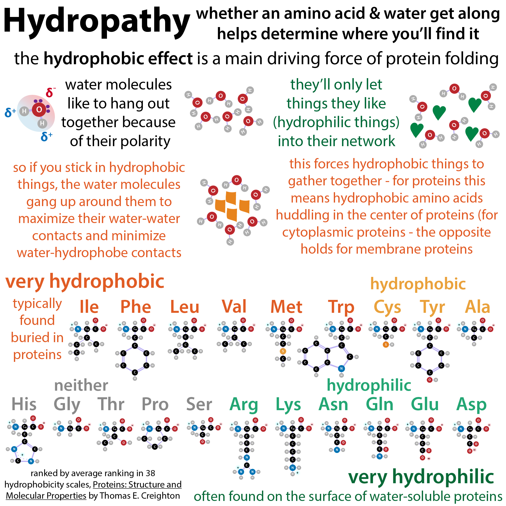
Alpha helixes are the most common way you’ll find proteins pass through. And, it takes ~20-30 amino acids per pass when they do! And not just any amino acids – you need nonpolar, hydrophobic ones so they’ll be happy hanging out with the hydrophobic lipid tails. Between each pass segment, however, as well as at exposed ends, you’ll need things to be hydrophilic so the watery exterior will play nice with them. It’s common for transmembrane proteins to have 7 or so passes.
It’s estimated that about 1/4 of our protein-coding genes (genes with instructions for making proteins) are recipes for integral membrane proteins³. And scientists can get this estimate by using the characteristic “signature” of alpha-helical transmembrane domains. They look for stretches of ~20 hydrophobic amino acids, which will show up as staple-shaped curves above the axis of a hydropathy plot*
*Scientists can classify amino acids by something called a hydropathy index which measures how unhappy proteins would be if you switched them from a nonpolar environment (such as the inside of a membrane) to a polar one (such as the cytosolic surface of the protein). Technical-wise, it measures the change in free energy (delta G) that occurs. More on free energy in other posts but positive is “unfavorable” so hydrophobic things will have a positive hydropathy index whereas hydrophilic things will have a negative one.
Beta barrels are harder to predict based on sequence alone because it only takes about 7 amino acids or so to span the membrane in the zig-zag arrangement of a beta strand, and they don’t have to all be hydrophobic. Instead, only every other amino acid will have to interact with a lipid (and thus be hydrophobic) whereas the others may form a hydrophilic pore that allows passage of hydrophilic things through the membrane. These pores are often made up of 20 or more strands and classic examples are porins such as the channels found in bacterial membranes.
References:
¹ Alberts B, Johnson A, Lewis J, et al. Molecular Biology of the Cell. 4th edition. New York: Garland Science; 2002. Membrane Proteins. Available from: https://www.ncbi.nlm.nih.gov/books/NBK26878/
² Fagerberg, L., Jonasson, K., von Heijne, G., Uhlén, M. and Berglund, L. (2010), Prediction of the human membrane proteome. Proteomics, 10: 1141-1149. https://doi.org/10.1002/pmic.200900258
Almén, M.S., Nordström, K.J., Fredriksson, R. et al. Mapping the human membrane proteome: a majority of the human membrane proteins can be classified according to function and evolutionary origin . BMC Biol 7, 50 (2009). https://doi.org/10.1186/1741-7007-7-50
³ Arinaminpathy, Y., Khurana, E., Engelman, D. M., & Gerstein, M. B. (2009). Computational analysis of membrane proteins: the largest class of drug targets. Drug discovery today, 14(23-24), 1130–1135. https://doi.org/10.1016/j.drudis.2009.08.006
Other articles:
Arinaminpathy, Y., Khurana, E., Engelman, D. M., & Gerstein, M. B. (2009). Computational analysis of membrane proteins: the largest class of drug targets. Drug discovery today, 14(23-24), 1130–1135. https://doi.org/10.1016/j.drudis.2009.08.006
Denisov, I., Sligar, S. Nanodiscs for structural and functional studies of membrane proteins. Nat Struct Mol Biol 23, 481–486 (2016). https://doi.org/10.1038/nsmb.3195
And if you have access to Lehninger’s Biochemistry textbook, chapter 11 has great stuff! Spent this morning rereading it.
As for past posts of mine that will provide more detail and/or background:
- more on water from a biochemistry perspective: https://bit.ly/dontignorewater & https://youtu.be/UQIHWIrhRu4
- more on the hydrophobic effect: http://bit.ly/hydrophobiceffectPSA & https://youtu.be/CJWEWrwUXI4
- more on soaps, detergents, & other amphiphiles: https://bit.ly/amphilphiles & https://youtu.be/kV3tPr7ZT4o
- more on protein structure: https://bit.ly/proteinstructure & https://youtu.be/V2pStHenOJo
- more on cryoEM: http://bit.ly/cryoemxray & https://youtu.be/KgrgVWbuypU
- more on the Novavax vaccine: https://bit.ly/novavaxvaccine & https://youtu.be/xmJX5t2g3KE
- more on receptors: https://bit.ly/receptorligands & https://youtu.be/Fg_uN-y5yak
- more on free energy & thermodynamics: http://bit.ly/partypopperscience & https://youtu.be/B2ifgzfZTtg