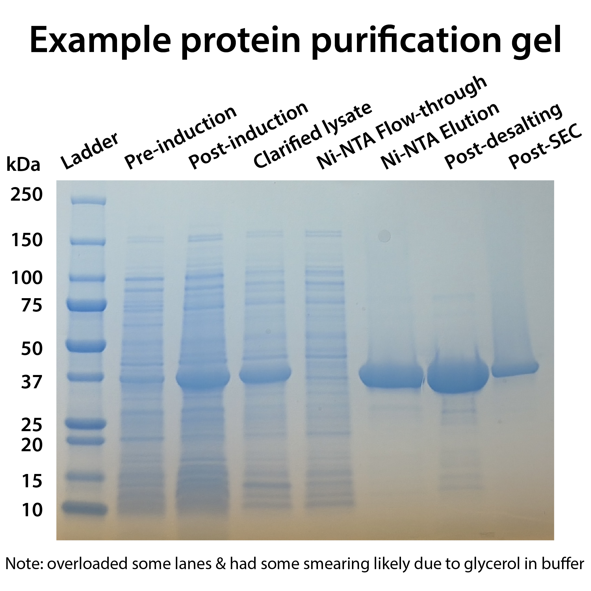
What can and can’t you tell just by looking at a gel? How about a blot? What can it tell you and what can it not?
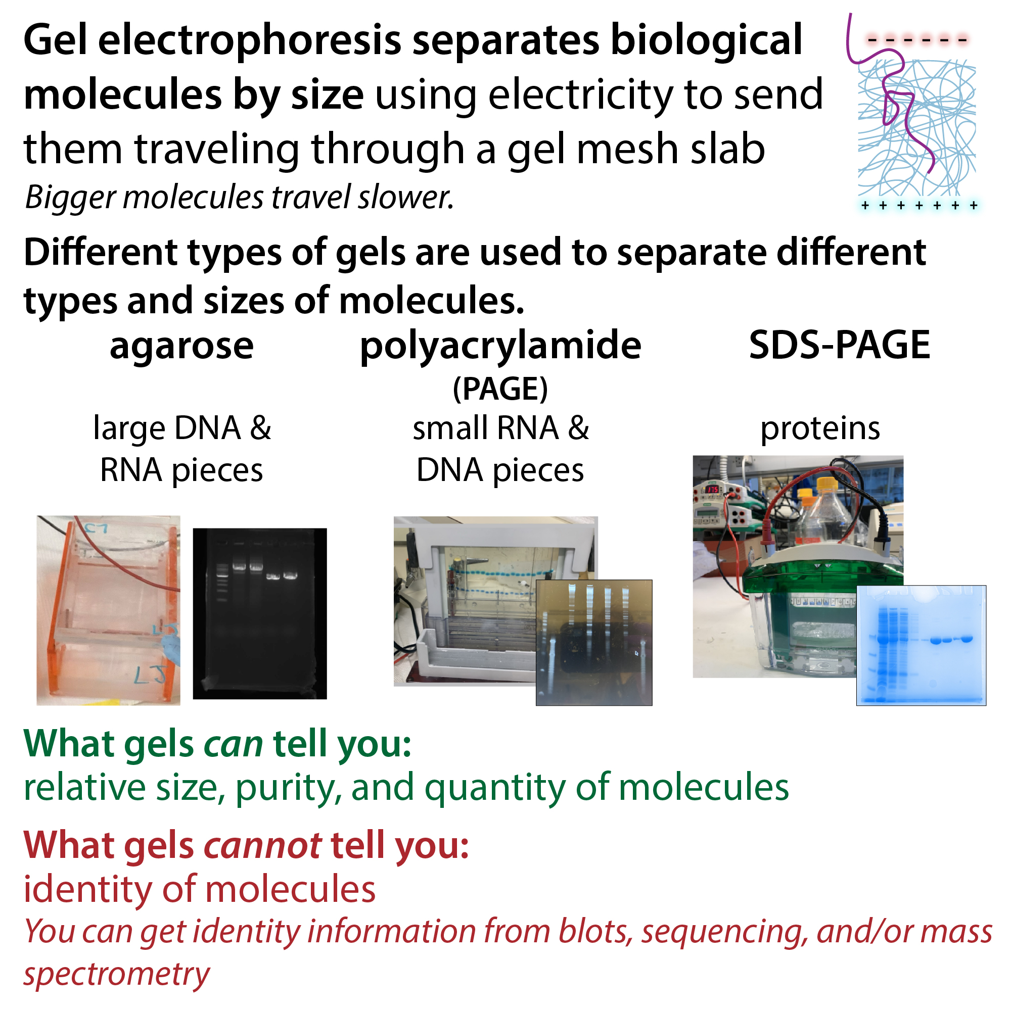
A simple protein, DNA, or RNA gel cannot tell you about the identity of the molecules present. It can’t really tell you anything conclusively (as you’ll see as I get deep into the caveats about overlapping bands and stuff).
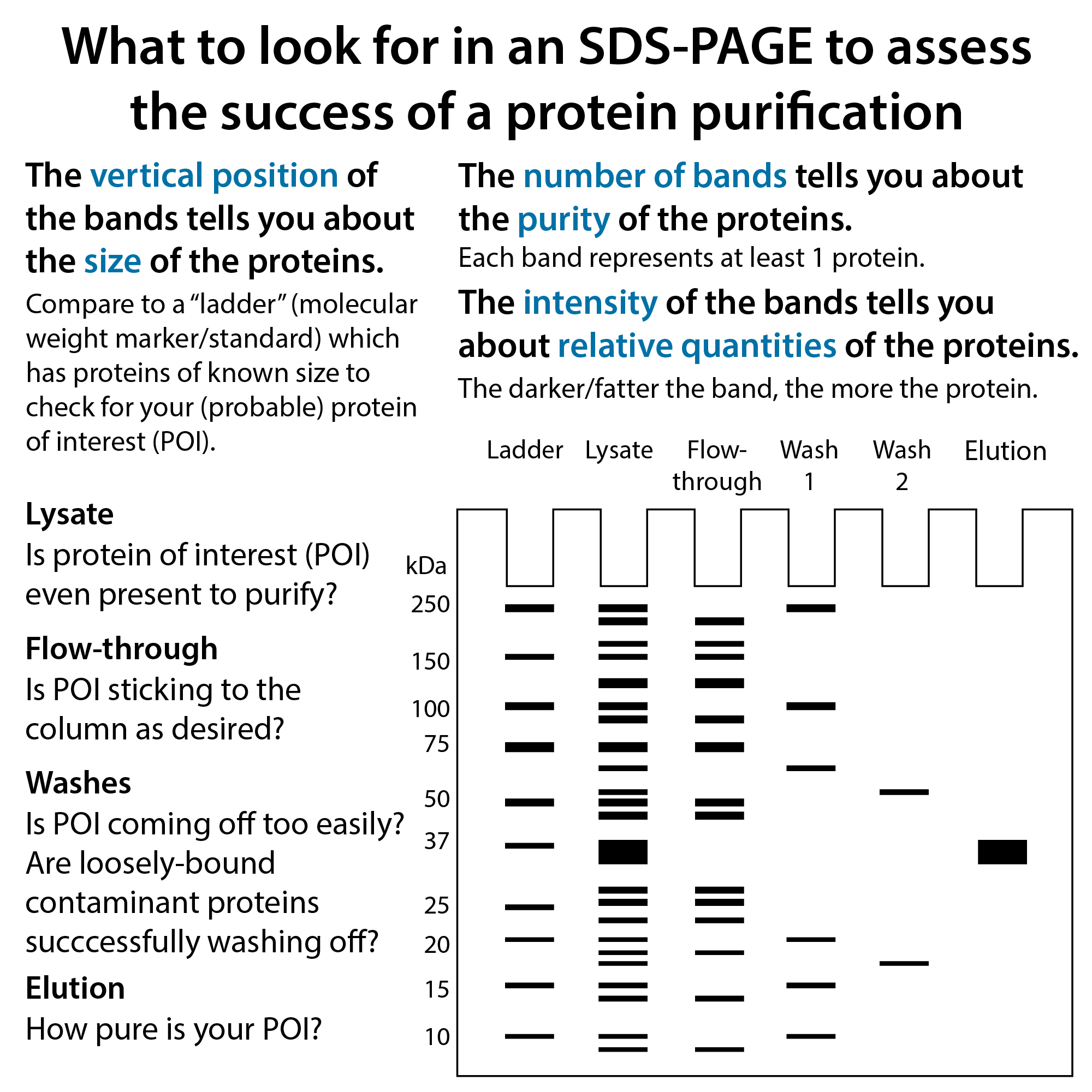
But it can tell you a rough idea about how many and how much of different molecules are present. If you are purifying or working with a purified protein, and you’re pretty sure that big fat band is your protein of interest, you can gauge how pure the protein really is – how many other bands are there? And what proportion of the total signal you see is the band that’s your protein of interest?
It can also tell you about the integrity of that molecule – do you see a bunch of smaller bands or a smear underneath it? This can indicate degradation – you might need to add protease or nuclease inhibitors or at least treat your samples with more TLC (and ice, avoid freeze-thaws, etc.)
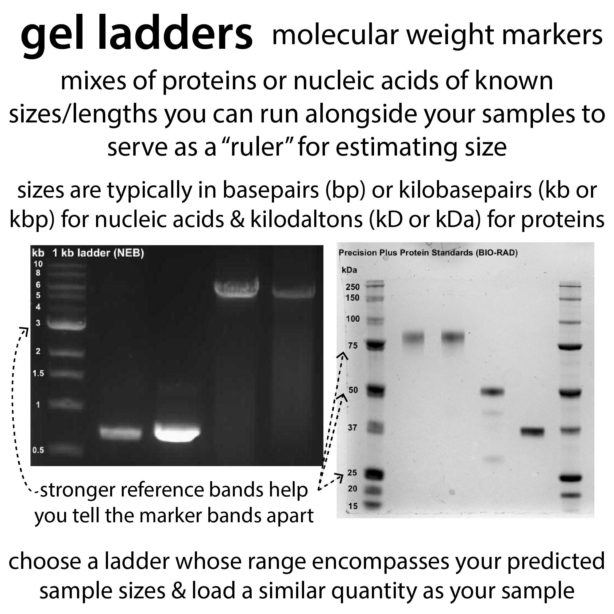
It can tell you about relative sizes of the different molecules present – you can compare to a “ladder” (molecular weight markers) you run alongside it. But beware that your protein might run slightly higher or lower than you’d expect based on its calculated molecular weight.
Also, if you see a kind of diffuse band, this could indicate a proportion of the molecules are modified (e.g proteins phosphorylated). But if it’s happening with multiple bands it could also just mean you ran the gel too fast or let it sit too long or loaded too much sample.
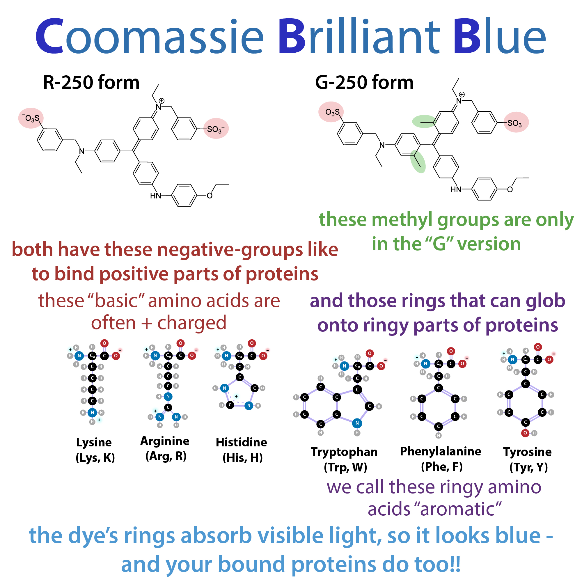
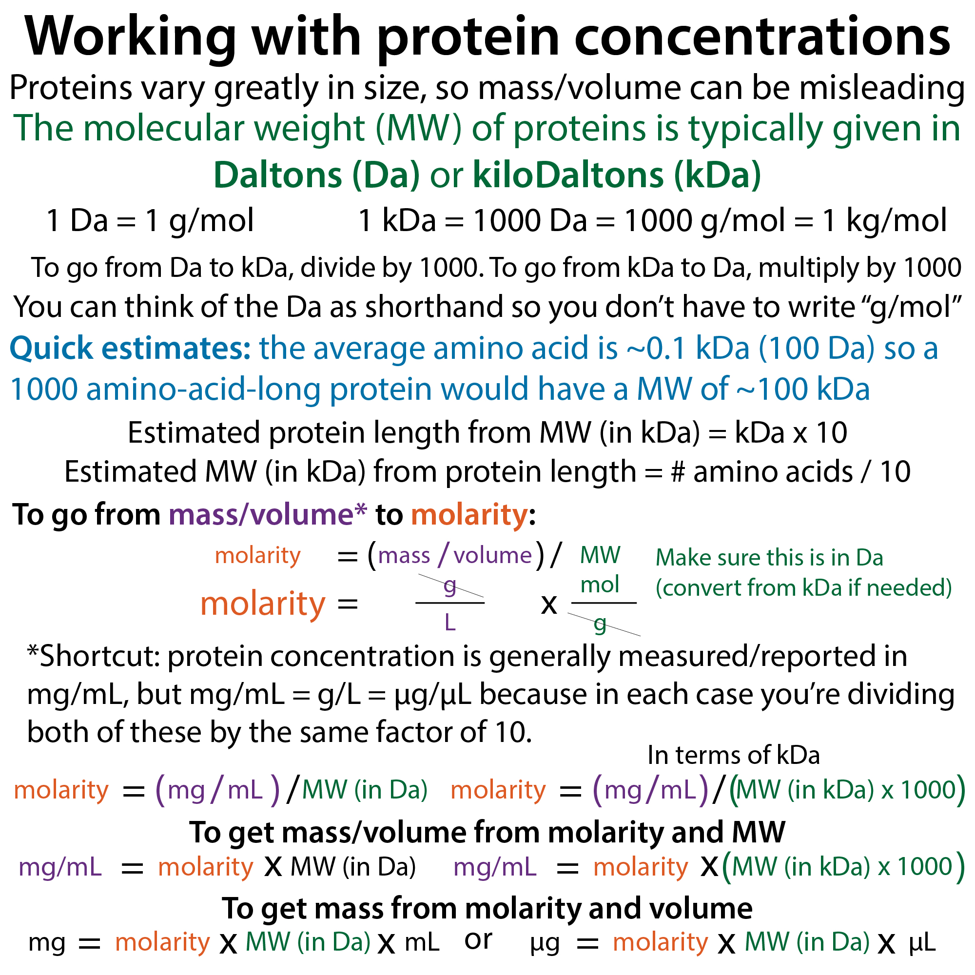
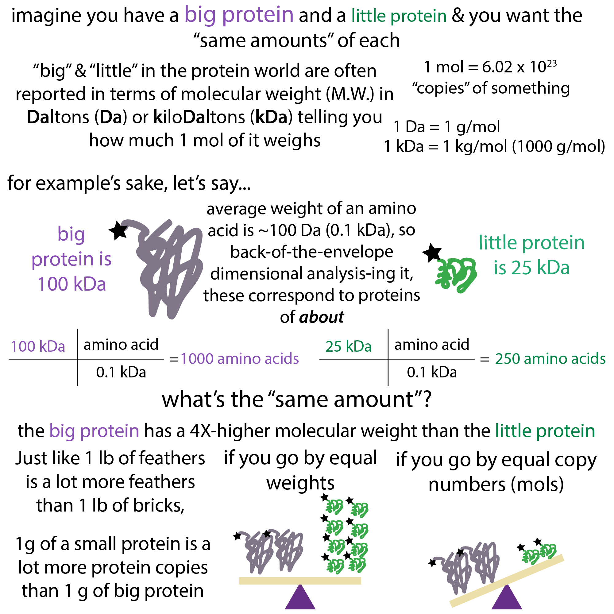
Band strength is connected to sample quantity, but it’s complicated. Because smaller molecules have less stuff for dye to bind to, you’ll need more copies of them to get the same strength as a bigger molecule. Additionally, and especially crucial with proteins, the composition of the protein can affect how well the dye binds to it (since these dyes typically prefer to bind certain amino acids and different proteins have different combos of amino acids, two proteins of the same size and quantity could have bands of different intensities).
Speaking of having multiple molecules of the same size – if you have multiple things of near the same size their bands will overlap so you might think you have a single band but really there are multiple. This is one reason why you want to choose your gel percentage to have the best “resolving power” in the range of your molecule of interest.
You can (often reasonably) predict that a band corresponds to a certain molecule based on its relative size and what you’re expecting to be there (especially in in vitro systems working with purified or partly-purified molecules). But in order to confirm the identity you’ll have to do something like a blot or mass spec or sequencing or something.
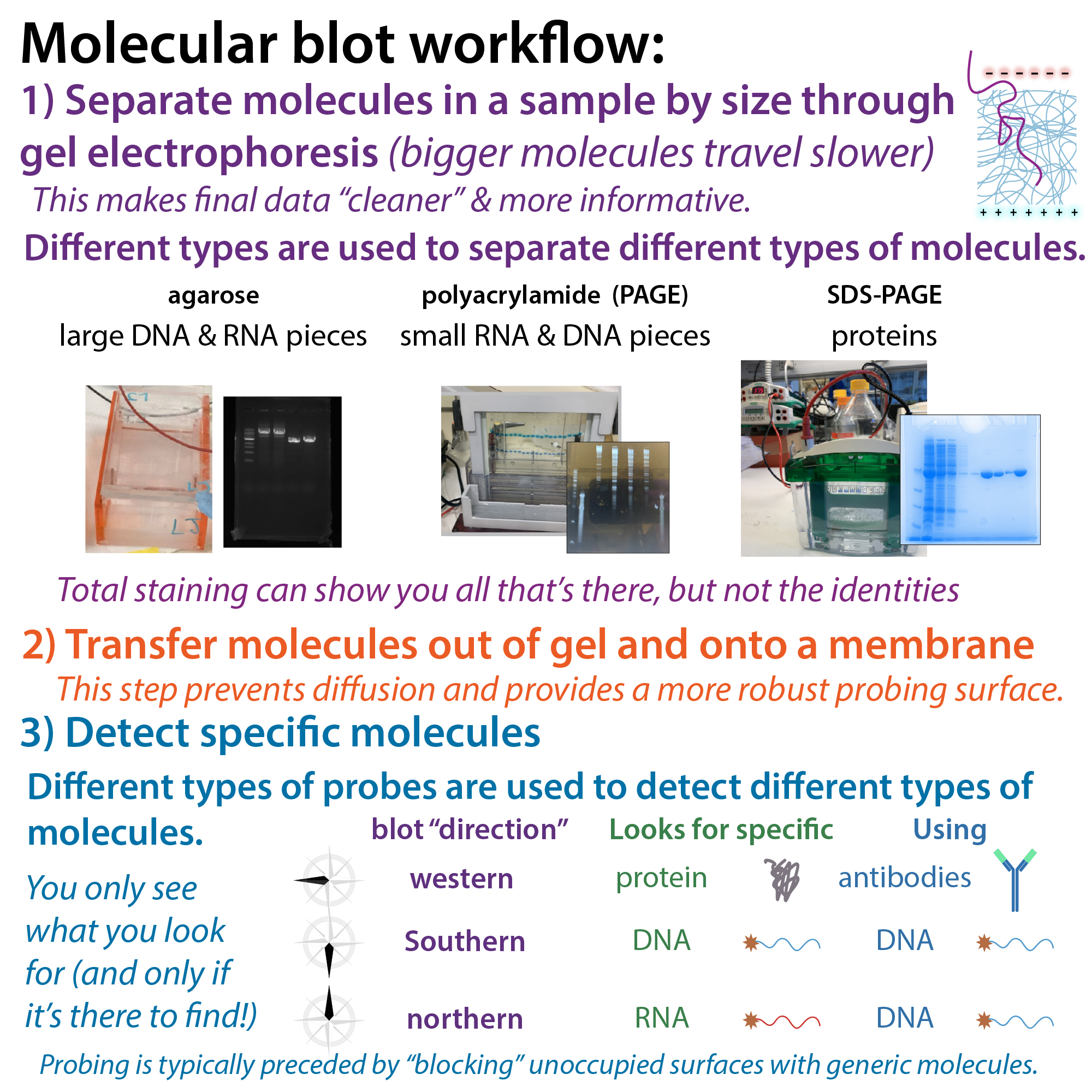
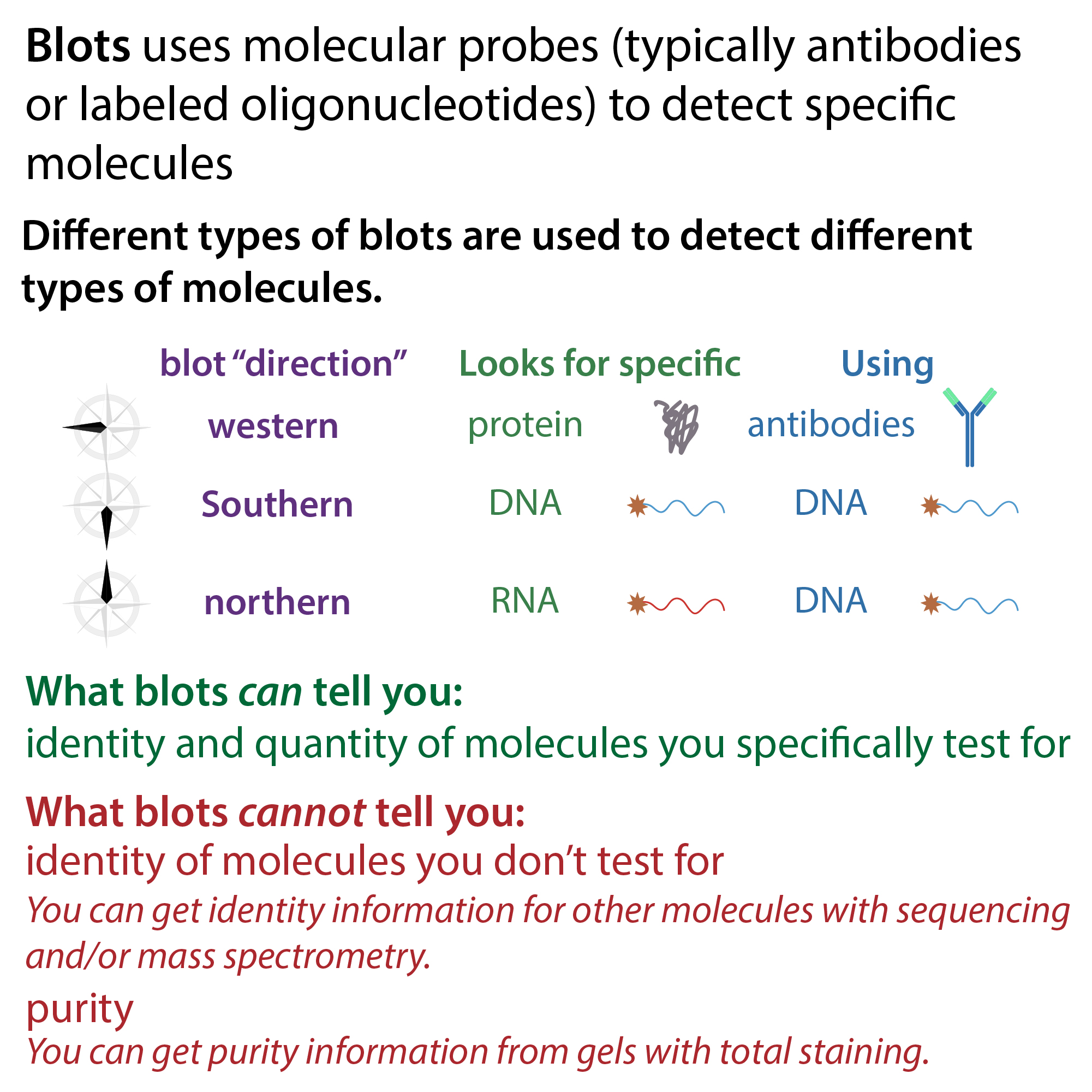
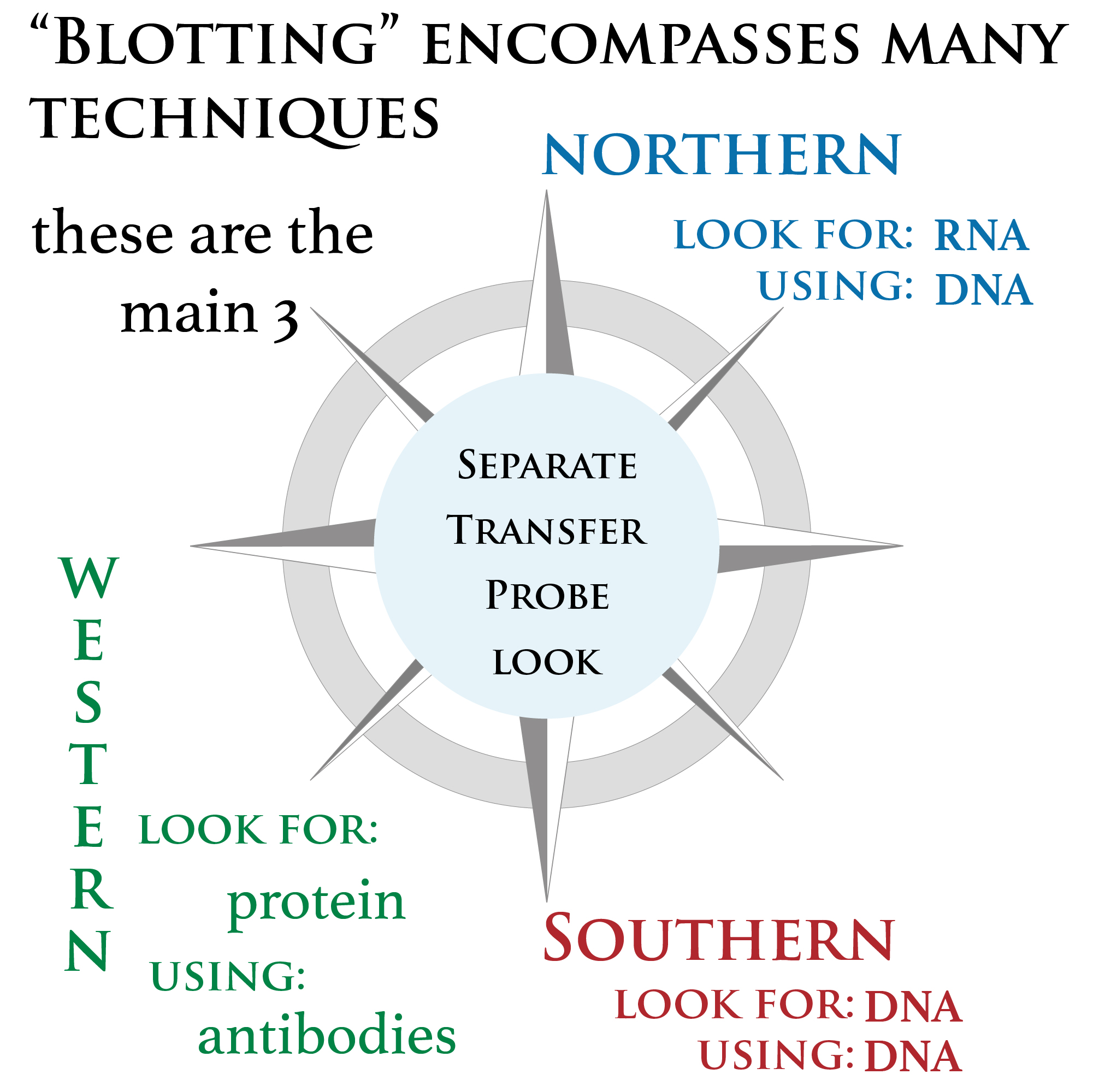
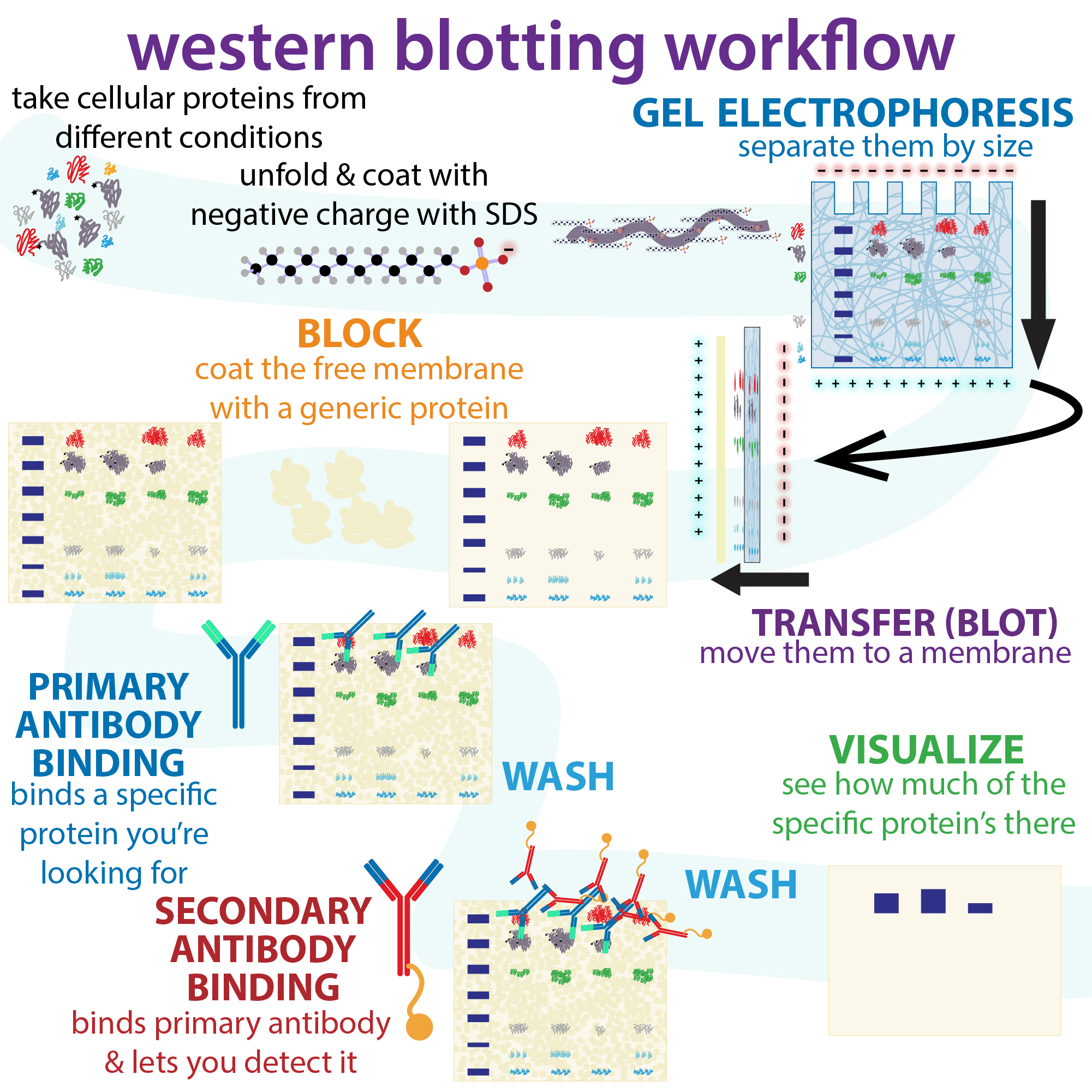
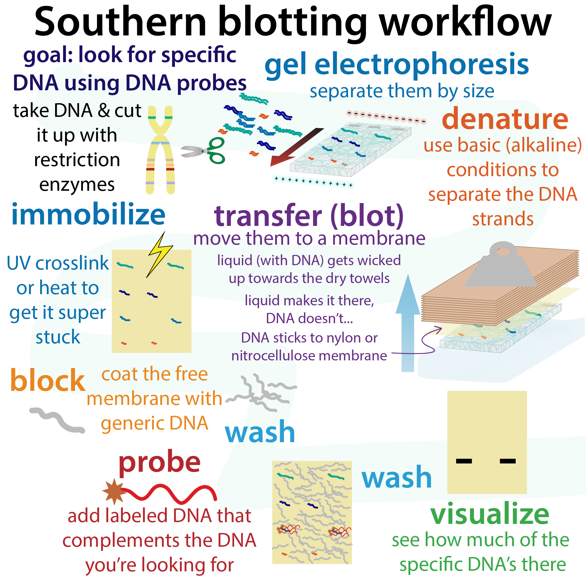
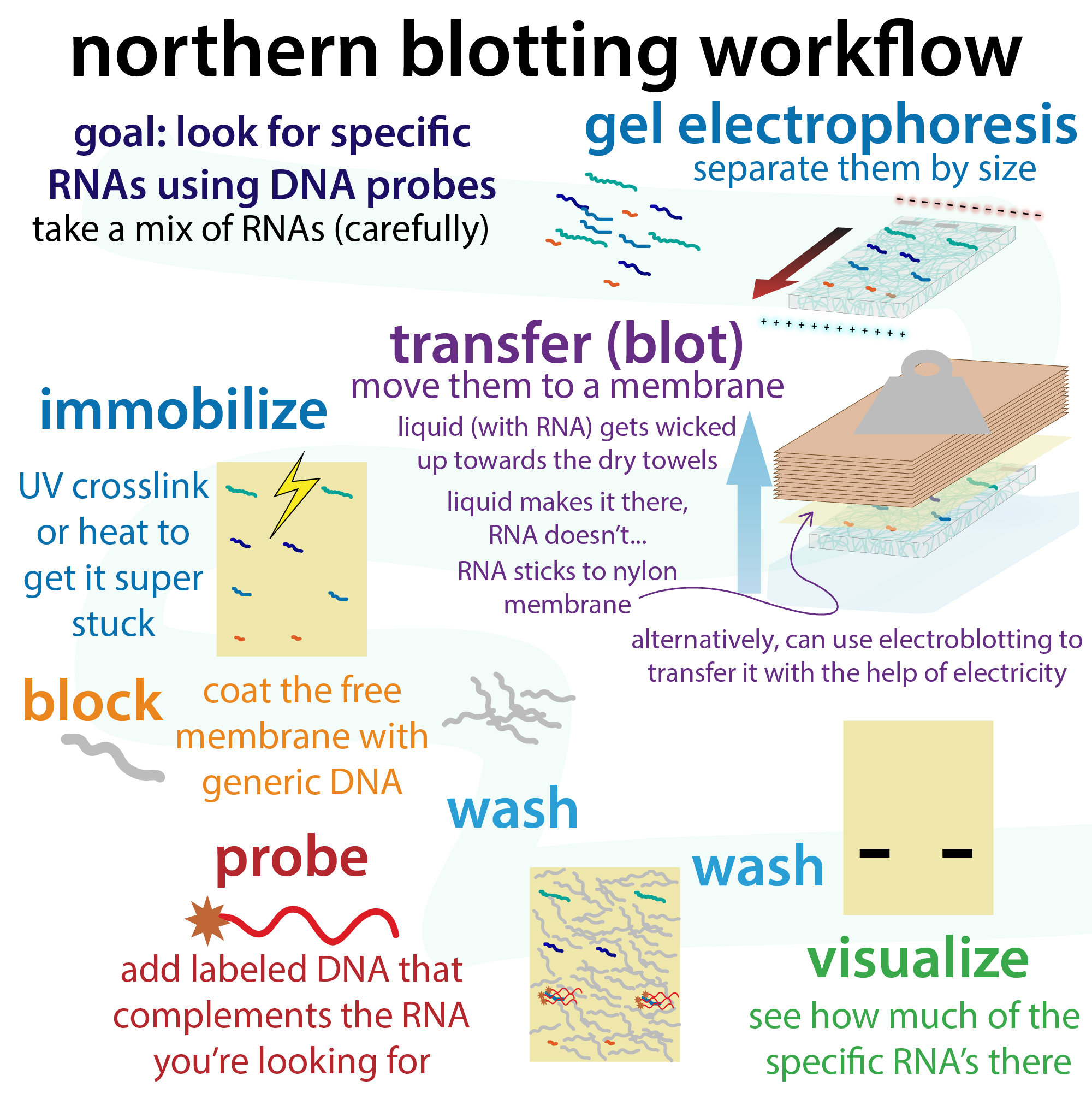
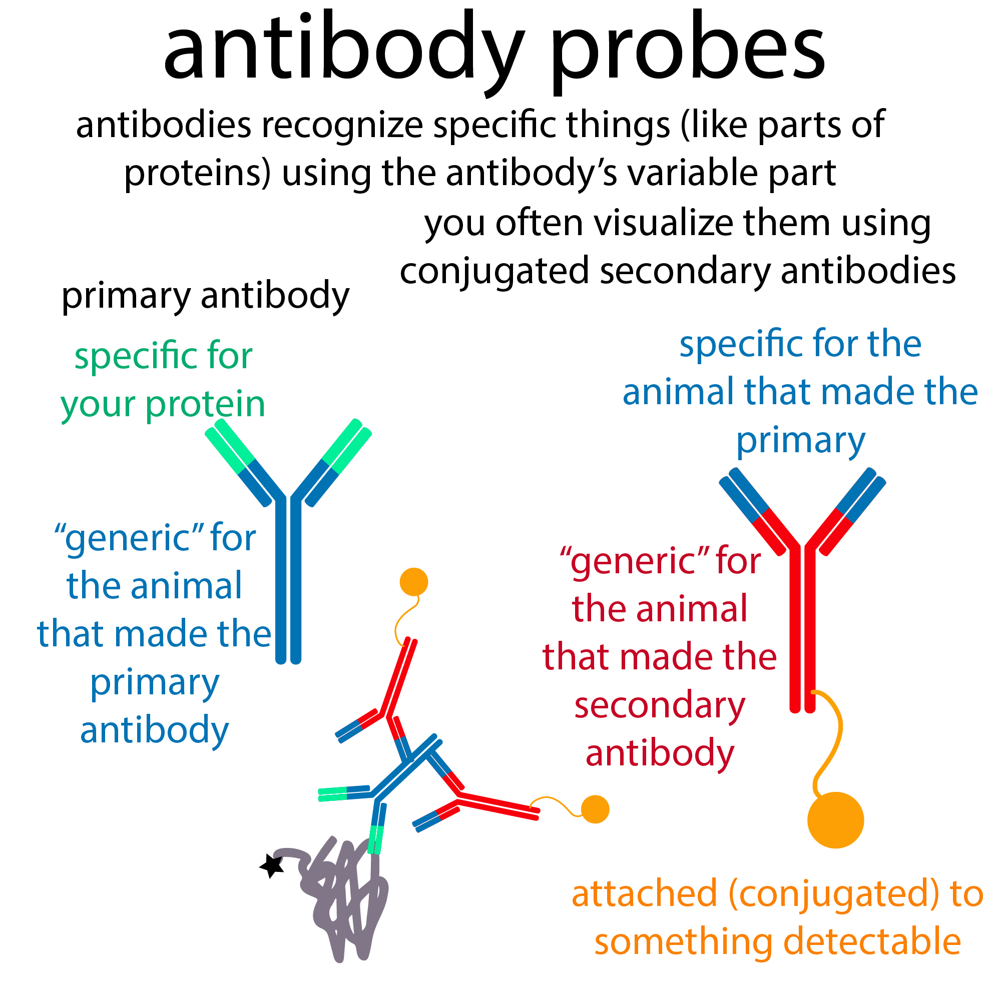
Blots use specific probes (antibodies for proteins in western blots and labeled DNA for DNA in Southern blots or RNA in norther blots) to look for specific molecules after transferring the molecules ouot of the gel and onto a membrane.
Blots aren’t perfect either though. They’re most useful for comparing relative amounts of a confirmed molecule in different samples. Different antibodies vary in strength. So if you have a really strong antibody you might see a stronger band for a protein that’s present in smaller amounts than the band you see for a protein with lots of it but poor antibodies. But if you compare the same protein, same antibody, but different samples, the fatter band indicates more’s there (when doing this to compare between samples, make sure you load equal “total sample” amounts).
Antibodies (in the protein case) and labeled oligonucleotide probes (for DNA and RNA) also vary in their specificity. Ideally they would only bind to the thing you want them to. Unfortunately they often also bind “non-specifically” to other things. So when possible use a negative control you know doesn’t have the thing you’re looking for (but has everything else) and a positive control of the purified thing you’re looking for so you know where the true band should be.
Blots tell you nothing about purity. So often all-protein or nucleic acid stains are also shown to tell you about that. These can be done separately (i.e. run 2 identical gels) or better-yet, they can be done by using a reversible stain on the membrane (such as a Ponceau stain for proteins).
So, in summary:
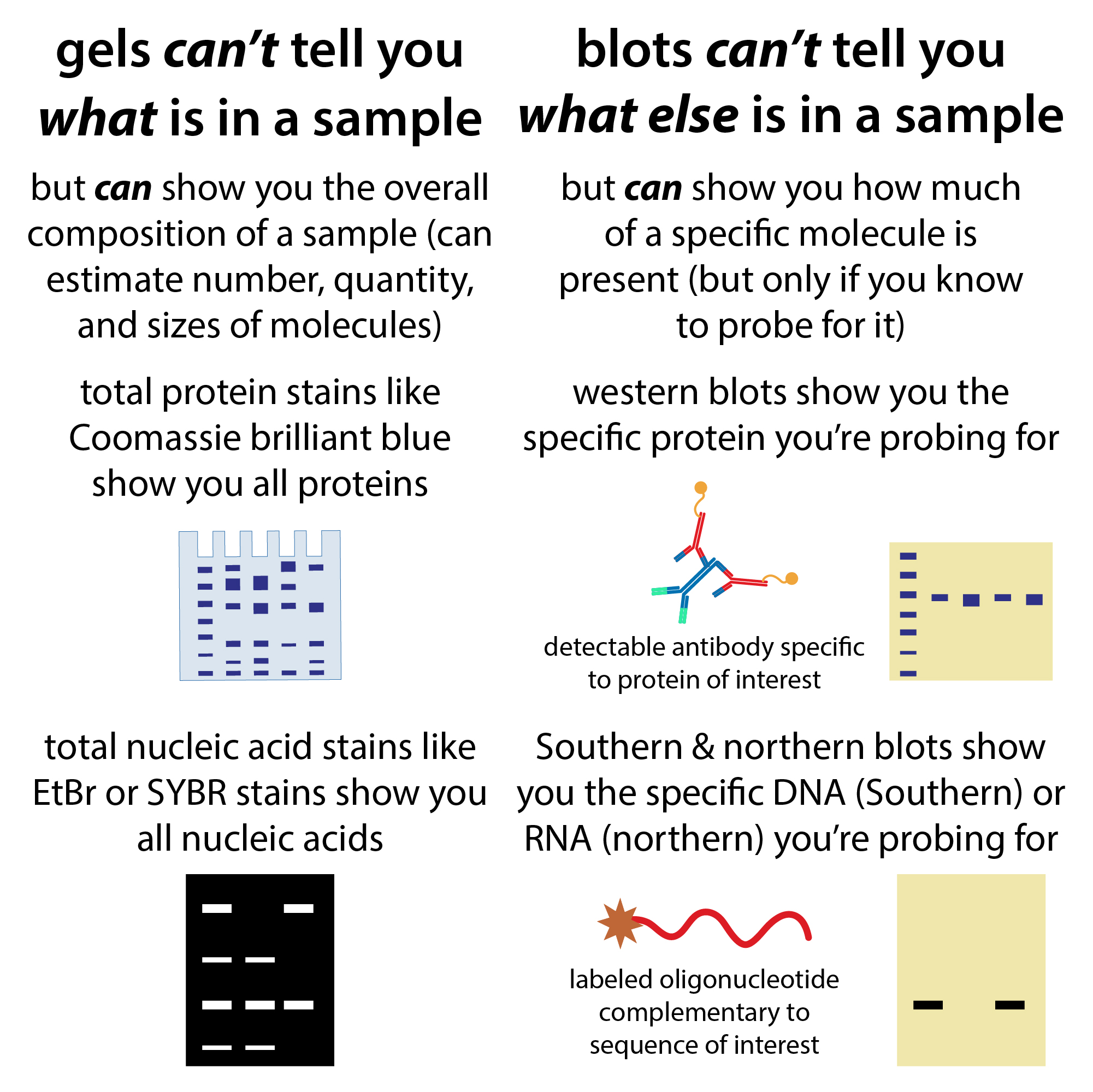
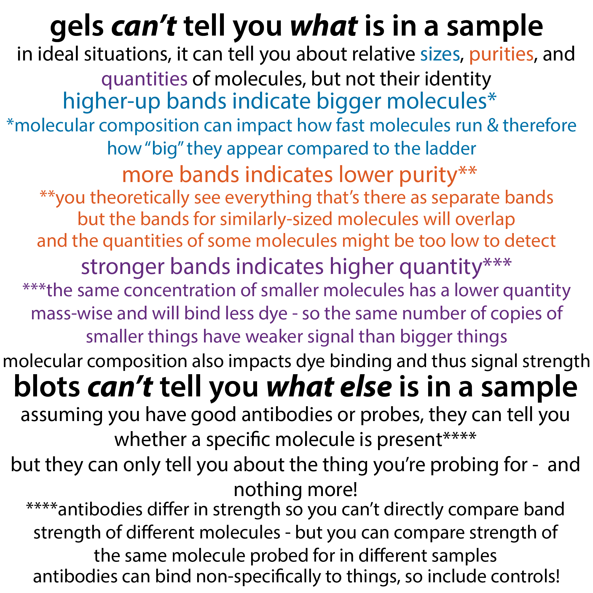
gels can’t tell you what is in a sample
- in ideal situations, it can tell you about relative sizes, purities, and quantities of molecules, but not their identity
- higher-up bands indicate bigger molecules*
- *molecular composition can impact how fast molecules run & therefore how”big” they appear compared to the ladder
- more bands indicates lower purity**
- **you theoretically see everything that’s there as separate bands, but the bands for similarly-sized molecules will overlap, and the quantities of some molecules might be too low to detect
- stronger bands indicates higher quantity***
- ***the same concentration of smaller molecules has a lower quantity mass-wise and will bind less dye – so the same number of copies of smaller things have weaker signal than bigger things
- molecular composition also impacts dye binding and thus signal strength
blots can’t tell you what else is in a sample
- assuming you have good antibodies or probes, they can tell you whether a specific molecule is present****
- but they can only tell you about the thing you’re probing for- and nothing more!
- ****antibodies differ in strength so you can’t directly compare band strength of different molecules – but you can compare strength of the same molecule probed for in different samples; also, antibodies can bind non-specifically to things, so include controls!
- similar holds true for nucleic acid probes – use longer probes and/or probes with higher Tms (stronger binding) for better specificity
more on blots: blog form (text old, video & some graphics new) : http://bit.ly/blotcompass; YouTube: https://youtu.be/dyWnkbVIOr4
more one western blots in particular: http://bit.ly/westernblotworkflow ; YouTube: https://youtu.be/Oun6_u6E090
more on gel electrophoresis: https://youtu.be/YoiNhxPNZJM & https://youtu.be/YoiNhxPNZJM
more on coomasssie staining: http://bit.ly/cbbgelstaining & https://youtu.be/LBsYSaZ6mBo
more on silver staining: https://bit.ly/silver_staining & https://youtu.be/B-e7dicwRtc
more on fluorescent nucleic acid stains: http://bit.ly/fluorescentstains & https://youtu.be/YoiNhxPNZJM
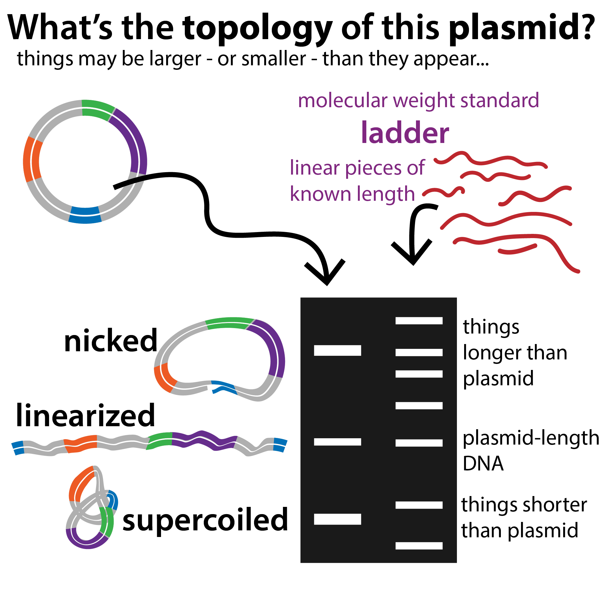
more on how DNA topology can affect how plasmids run: http://bit.ly/DNAtopology & https://youtu.be/4dK13lmZd8k
more on protein purification: http://bit.ly/proteinpurificationtech