Buffer you load a gel you have to mix it with a sample loading buffer! These buffers have a few key purposes (and thus necessary components)
- Brighten things up by bringing some color!
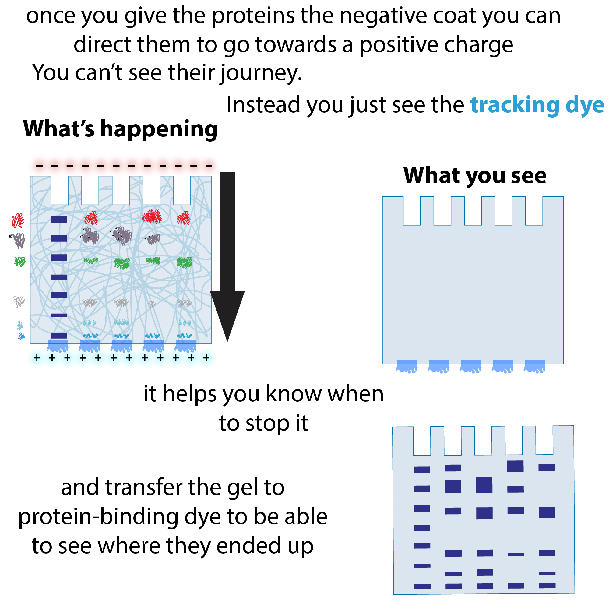
- A tracking dye (bromophenol blue, xylene cyanol, amaranth, etc.) lets you see your sample as you load it and, more importantly, track the progress of the run – can give you a heads-up if there’s a problem and also lets you know when to turn off the electricity to stop the run (before your samples run all the way out!)
- Warning: the bands you see are just the dye front – it is *not* showing you the molecules you are looking for (proteins, nucleic acids, etc.). You (usually) have to post-stain to see those bands
- A tracking dye (bromophenol blue, xylene cyanol, amaranth, etc.) lets you see your sample as you load it and, more importantly, track the progress of the run – can give you a heads-up if there’s a problem and also lets you know when to turn off the electricity to stop the run (before your samples run all the way out!)
- But weigh your samples down!
- Something heavy (glycerol, Ficoll 400, formamide (for denaturing nucleic acid buffers) to make your sample sink to the bottom of the well and stay there while you load all the wells
- Warning: though heavy and viscous, these guys are no match for the power of ethanol to pop your sample right out! So make sure you get all the ethanol out when doing nucleic acid purifications!
- Something heavy (glycerol, Ficoll 400, formamide (for denaturing nucleic acid buffers) to make your sample sink to the bottom of the well and stay there while you load all the wells
- Denaturant? (SDS, formamide, etc.)
- pH buffer? (Tris, etc.)
- EDTA?
YouTube: https://youtu.be/Q52IqEuhXKU
The dye and heavy thing are the only *required* components for a non-denaturing (aka native) gel – but you often want to denature as well!
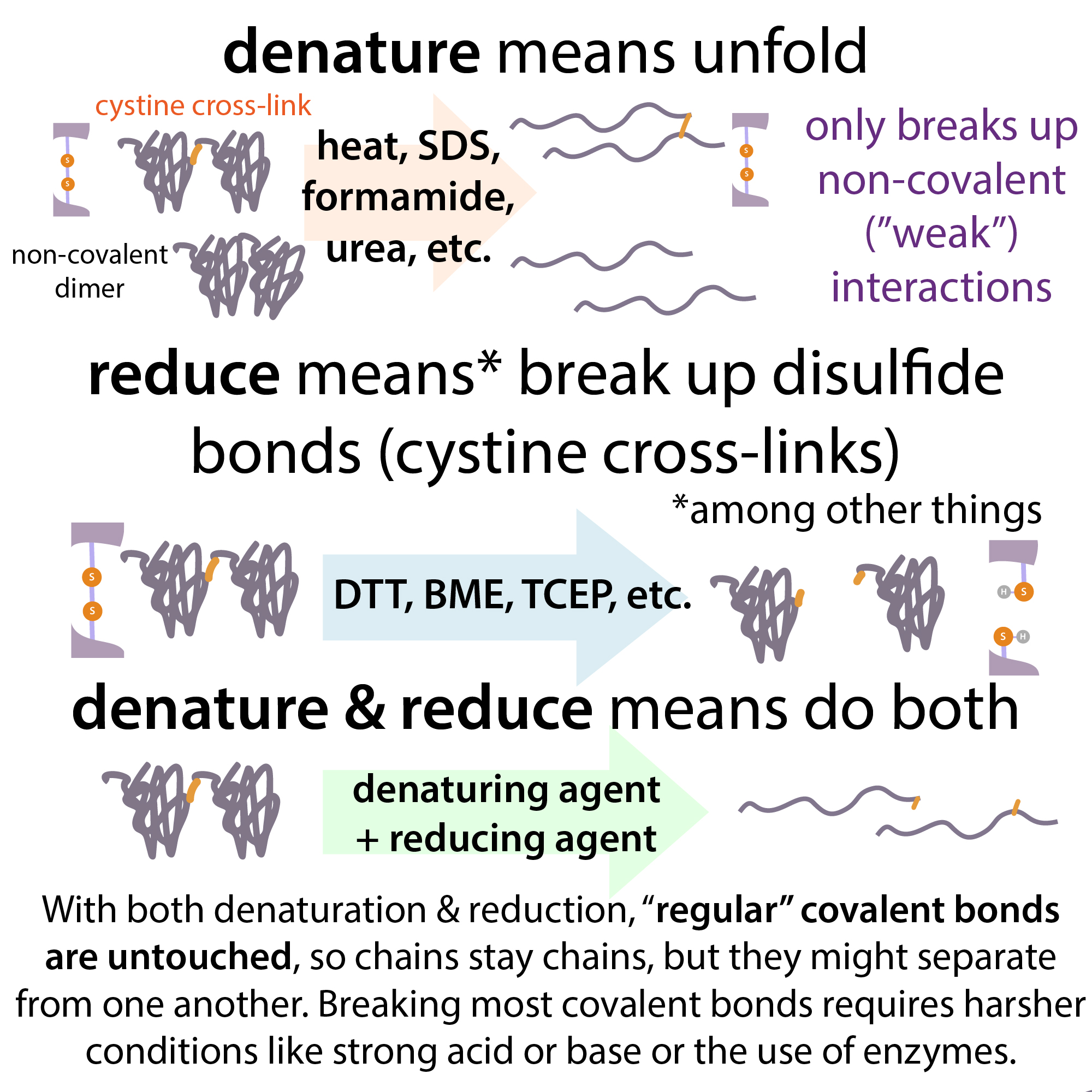
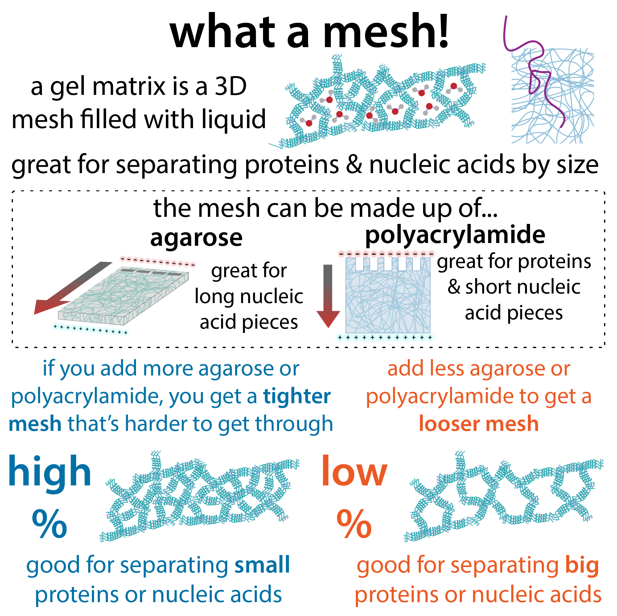
Denaturing refers to unfolding molecules to remove their shape and breaking up intermolecular (between-molecule) interactions. We often want to denature our samples before we run them so that their shape doesn’t get in the way of their slithering through the gel mesh. This way, they will travel at a speed “only” proportional to their linear length.
We use different denaturants for proteins and nucleic acids. For proteins, we usually use SDS (sodium dodecyl sulfate), whereas we usually use formamide for nucleic acids. In both cases, we typically use them in combination with heat – the heat helps get the denaturation going and then these guys bind to and help stabilize the unfolded molecules.
SDS is a detergent (artificial soap) that has a hydrophobic (water-avoided) tail and a hydrophilic (water-loved) head. Those hydrophobic parts glob onto the exposed hydrophobic parts of the protein (that had been hidden at the protein’s core) while the heads help keep things soluble. And the heads are negatively-charged (anionic), which provides the even negative charge needed to motivate the proteins to move through the gel towards the positive charge the electrophoresis box sets up.
For nucleic acids, we don’t have to worry about adding a negative charge, since the phosphodiester backbone of DNA and RNA does a great job of that for us. But we may want to unfold RNA secondary structure (things like hairpins that form when the RNA folds back on itself) or un-double-strand (melt) double-stranded DNA. For this, formamide provides a way. This molecule disrupts the intermolecular hydrogen bonds that had been holding those things together, replacing them with bonds to itself. To prevent the molecules from refolding, rebinding, etc during the run, we usually include another denaturant, urea, in the gel itself which can take over if the formamide falls off.
In the case of proteins, if you really want to get everything all linear and monomeric (each protein chain separate) you should add a reducing agent as well. Commonly this is BME (beta-mercaptoethanol) or DTT (dithiothreitol) and what it does is it breaks of disulfide crosslinks which are too strong for SDS to disrupt but weaker than the rest of the bonds holding amino acids together within an individual chain. Because these reducing agents go bad over time (they get oxidized by things other than our crosslinks so they can no longer reduce our crosslinks) we usually add them fresh and store sample buffer containing them in the -20 freezer.
A couple other things you might find in the buffer
- an actual buffer! You often see Tris in SDS-PAGE buffers, which helps stabilize the pH where you want it
- Nucleic acid sample buffers often have EDTA which is a “chelator” meaning it bites down on metals, thus hiding them from nucleases (DNA and RNA chewers). It also can help hide metals that might otherwise stick to the nucleic acids and interfere with how they run
A couple final notes:
It’s good to keep an eye on the dye front from the start (not just when you think it should be close to done) – this lets you see if there are any problems. What you want to see is that the dye front is moving evenly (all the lanes at the same rate). If you see one side running higher than the other, this is sometimes because the buffer level is too low (is your cassette leaking? If so, once your samples are in the gel (cleared past the wells) you can take it out and fix it) or you might see a smiling effect where the edges run slower than the center due to uneven heating. If you see that, turn down the voltage. The sooner you can detect a problem, the sooner you can try to fix it. more here: blog: https://bit.ly/gel_smile ; YouTube: https://youtu.be/a8geIlRuwrc
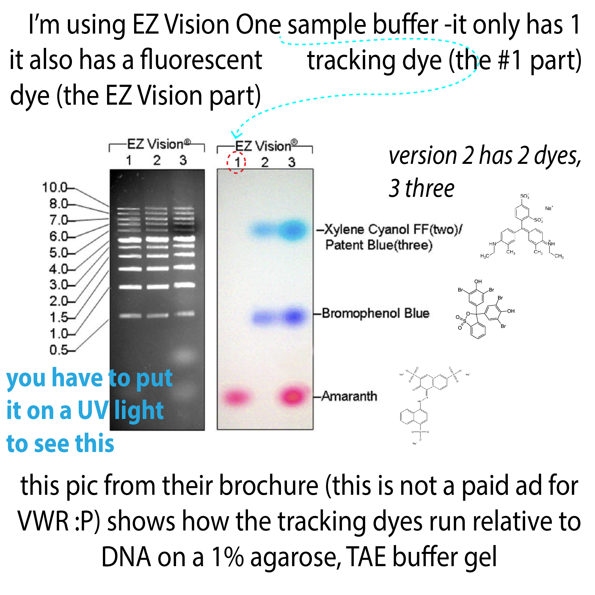
In terms of the dyes in nucleic acid sample loading buffers, you often see 2 or 3. This allows you to estimate where various size fragments are in the gel during the run. The dyes run differently in different gel types and percentages, so check a dye migration chart to see what size fragments the dyes will run alongside in the gel you’re using. If they overlap with yours they could make it hard to see your bands so you may want to use another dye. You may need to make one yourself but it’s super pretty to do so!
Here are a few I use:
SDS-PAGE (denaturing protein):
The recipe we use in our lab for 100mL of 6X is:
- 5.91 g Tris-HCl pH 6.8
- 6 g SDS
- 48 mL glycerol
- 30 mg bromophenol blue
You can store this at room temperature (RT). Then, before using, add 90 μL of β-mercaptoethanol to 910 μL of this mix – you can freeze this aliquot and it will be good for a while. Note: many protocols use higher [BME] than this (5% is common, but our recipe uses 1.5…)
working concentrations:
- 80 mM Tris-HCl pH 6.8
- 1% SDS (w/v)
- 8% glycerol (v/v)
- 0.0005% bromophenol blue (w/v)
non-denaturing nucleic acid:
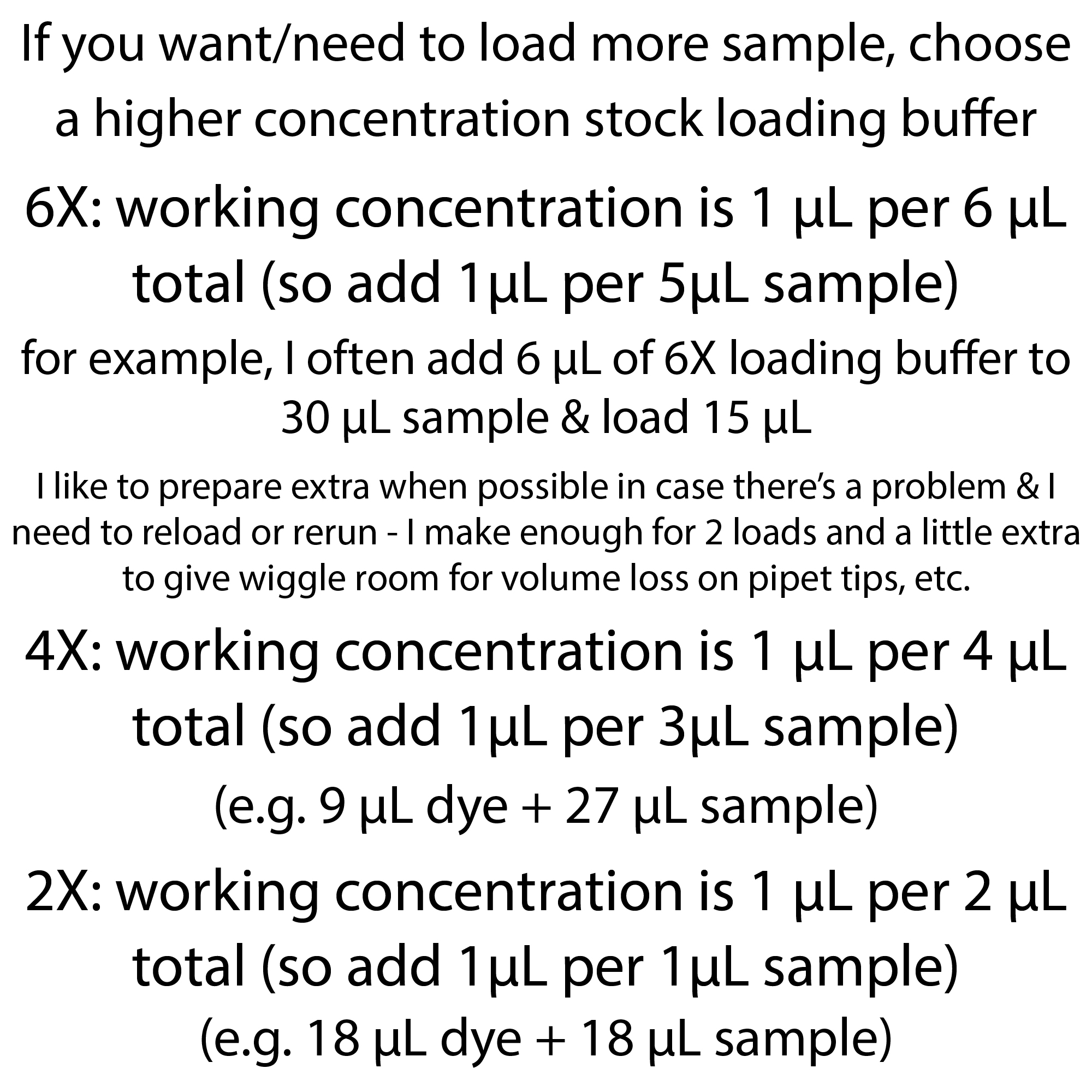
- 6X Non-denaturing sample loading buffer (6X NSLB): 10mM Tris pH 8; 1mM EDTA; 15% w/v Ficoll 400; 0.25% w/v Bromophenol Blue
denaturing nucleic acid:
- 2X Denaturing Sample Loading Buffer (2X DSLB): 98% v/v formamide; 10mM EDTA; 300μg/mL bromophenol blue
- 2X Xylene cyanol formamide loading dye: 95% formamide, 5mM EDTA (pH 8.0), 0.05% (w/v) xylene cyanol
for more on SDS-PAGE loading buffers: blog: https://bit.ly/sds_loading_dyes ; YouTube: https://youtu.be/IDENwOYpGRs
more on denaturing vs. reducing https://bit.ly/denaturing_vs_reducing ; YouTube: https://youtu.be/YoMH7CmYHwA
more on SDS-PAGE: http://bit.ly/sdspageruler & https://youtu.be/FgXlPAKVSGY
more on native protein-PAGE: https://bit.ly/nativepageoverview & https://youtu.be/QRL5cfJNwm0
more on nucleic acid PAGE: http://bit.ly/ureapage & https://youtu.be/MHJqnur6yqk
more about agarose gel electrophoresis: http://bit.ly/agarosegelelectrophoresis & https://bit.ly/agarosegelcompare & https://youtu.be/vbuxf3rcMxg
more about all sorts of things: #365DaysOfScience All (with topics listed) 👉 http://bit.ly/2OllAB0 or search blog: https://thebumblingbiochemist.com