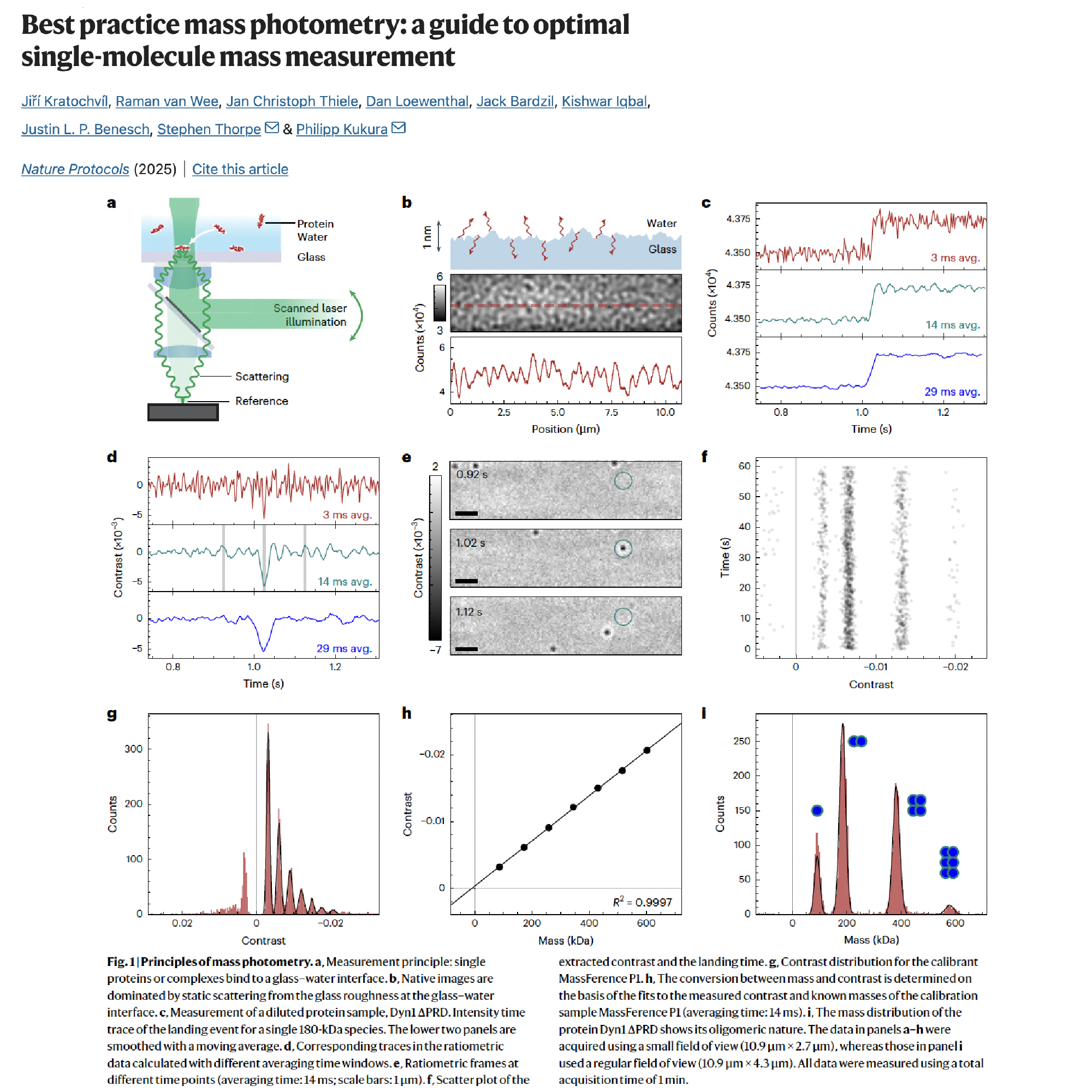
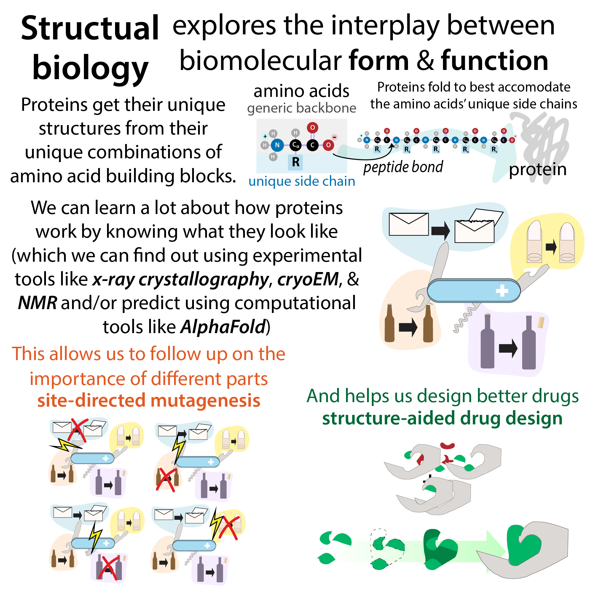
Mass photometry (MP) is a cool technique that you can use to determine the mass as well as the distribution of molecules of different masses in a solution. This allows you to do things like figure out if a solution is pure determine the relative memorization of a complex see if store chemic stoichiometry is correct etc. It’s growing in use, especially in the structural biology field where it can be used to screen samples under different conditions and things before going to do something like cryo-EM or x-ray crystallography where you wanna make sure that you have a very pure, homogenous mixture.
The way it works is cool too. You basically have a glass slide and you shine light at it and you have a very dilute solution of your proteins and as they come into contact with the slide, they’re kind of transiently attached to it, and when they attached to it now they’re kind of gonna block the light that gets reflected from the shot slide from below. The more massive the molecule, the darker the sort of shadow will be on the detector so you detect a larger difference in contrast. The technique measures each molecule at a time over time, and so it makes single-molecule measurements unless that’s why it needs to be really dilute but rather than just measuring a single molecule you’re gonna be of molecules and then you tally up how many times you count molecules of each difference contrast level, which you display as a histogram. And then you can use a calibration curve where you have a protein of known sizes that then you can use that in order to figure out the relative size of your protein. And then, based on how many different size things you see in there you’ll know if your thing is pure and based on how many times you see something with that intensity you can determine relative amounts of molecules (how much tetramer vs. dimer, etc.)
It works for molecules 30 kDa to 5 MDa at 100 pM – 100 nM concentrations, which is really low, so you have to have tight binding in order for this to work but if it works then that’s cool. It’s not so cool is that you have to have a special machine in order to do this technique. This technique was only introduced in 2018 so it’s still fairly new so hopefully the cost of equipment will come down. I only learned about it fairly recently and so that’s why I don’t have much information on it and why it’s not used or known about as much but here are some articles that can help you learn more as well as a really cool webinar.
Unlike some techniques like SEC-SAXS (size exclusion chromatography-small angle light scattering) you’re not giving any sort of shape information just the molecular weight but you don’t need to have a beamline or something like you would to generate the x-rays needed for SEC-SAXS. You just have to have one of these optometry devices which I wish I had, but thankfully, I have collaborators.
Resources
Original paper: Quantitative mass imaging of single biological macromolecules | Science. https://www-science-org.lmu.idm.oclc.org/doi/10.1126/science.aar5839 (accessed 2026-03-14).
Highlight of original paper: Lee, S. F.; Klenerman, D. Weighing One Protein with Light. Science 2018, 360 (6387), 378–379. https://doi.org/10.1126/science.aat5851.
Uses in structural biology: Vostal, L. E. Need for Speed: Mass Photometry as a Sample Analysis Tool for Structural Studies. Structure 2025, 33 (6), 994–996. https://doi.org/10.1016/j.str.2025.04.014.
Uses in interaction studies: Kofinova, Z.; Karunanithy, G.; Ferreira, A. S.; Struwe, W. B. Measuring Protein-Protein Interactions and Quantifying Their Dissociation Constants with Mass Photometry. Current Protocols 2024, 4 (1), e962. https://doi.org/10.1002/cpz1.962.
Protocol: Kratochvíl, J.; van Wee, R.; Thiele, J. C.; Loewenthal, D.; Bardzil, J.; Iqbal, K.; Benesch, J. L. P.; Thorpe, S.; Kukura, P. Best Practice Mass Photometry: A Guide to Optimal Single-Molecule Mass Measurement. Nat Protoc 2025, 1–25. https://doi.org/10.1038/s41596-025-01255-4.
Webinar: Mass Photometry, The Protein Society, 1/5/26: https://youtu.be/3eLqRyviTII?si=tKX1zPqWSqePOKi_
SEC-SAXS: Putnam, C. D.; Hammel, M.; Hura, G. L.; Tainer, J. A. X-Ray Solution Scattering (SAXS) Combined with Crystallography and Computation: Defining Accurate Macromolecular Structures, Conformations and Assemblies in Solution | Quarterly Reviews of Biophysics. Cambridge Core 2007. https://doi.org/10.1017/S0033583507004635.
much more structural biology content: https://bit.ly/structural_biology & https://youtube.com/playlist?list=PLUWsCDtjESrGhwVxsRbTJdL-BEsN60RCs