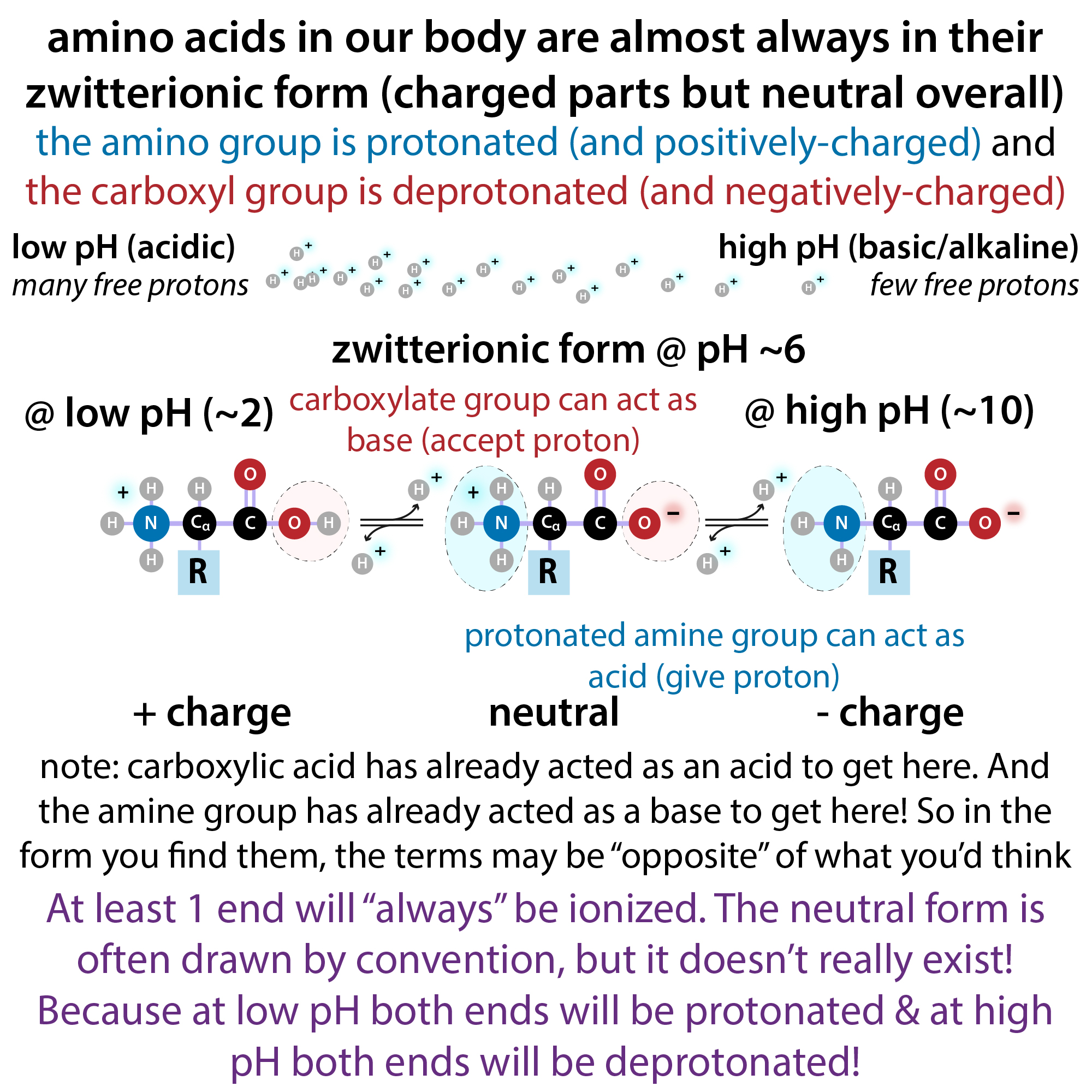
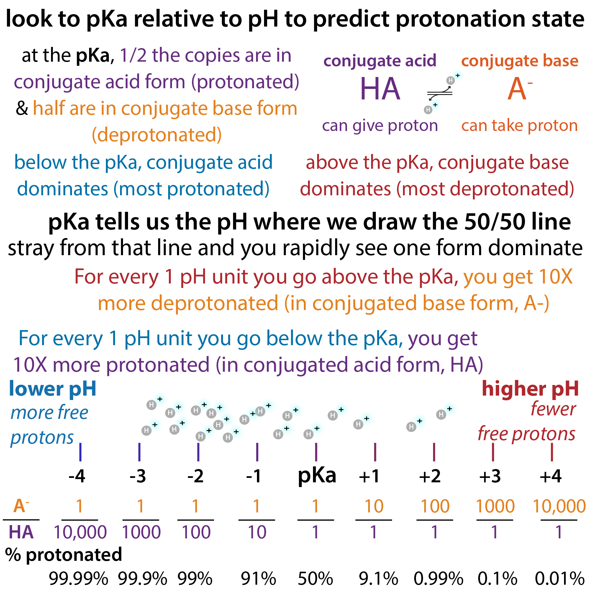
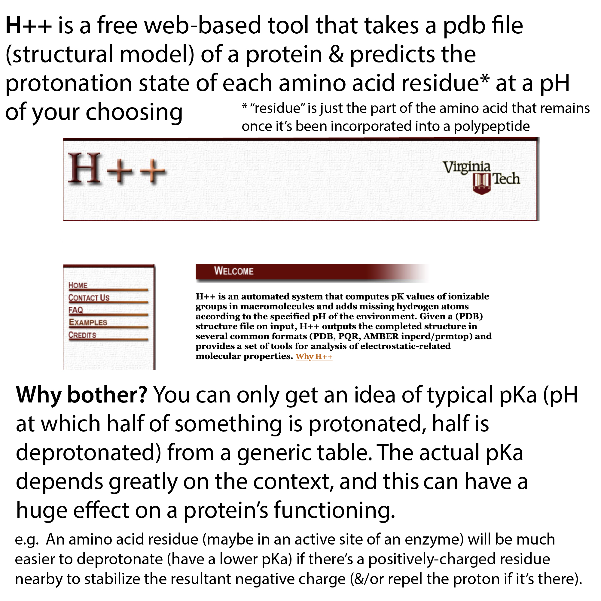
We’ve noticed some pH-dependent changes in our protein, so a free web-based tool that I’ve been exploring is H++. It takes a pdb file (structural model) of a protein and predicts the protonation state of each amino acid residue at a pH of your choosing. This is important, because you can really only get an idea of typical pKa (pH at which half of something is protonated, half is deprotonated) from a generic table. The actual pKa depends greatly on the context. For example, an amino acid residue will be much easier to deprotonate (have a lower pKa) if there’s a positively-charged residue nearby to stabilize the resultant negative charge (and/or repel the proton if it’s there). And this can have a huge effect on a protein’s functioning.
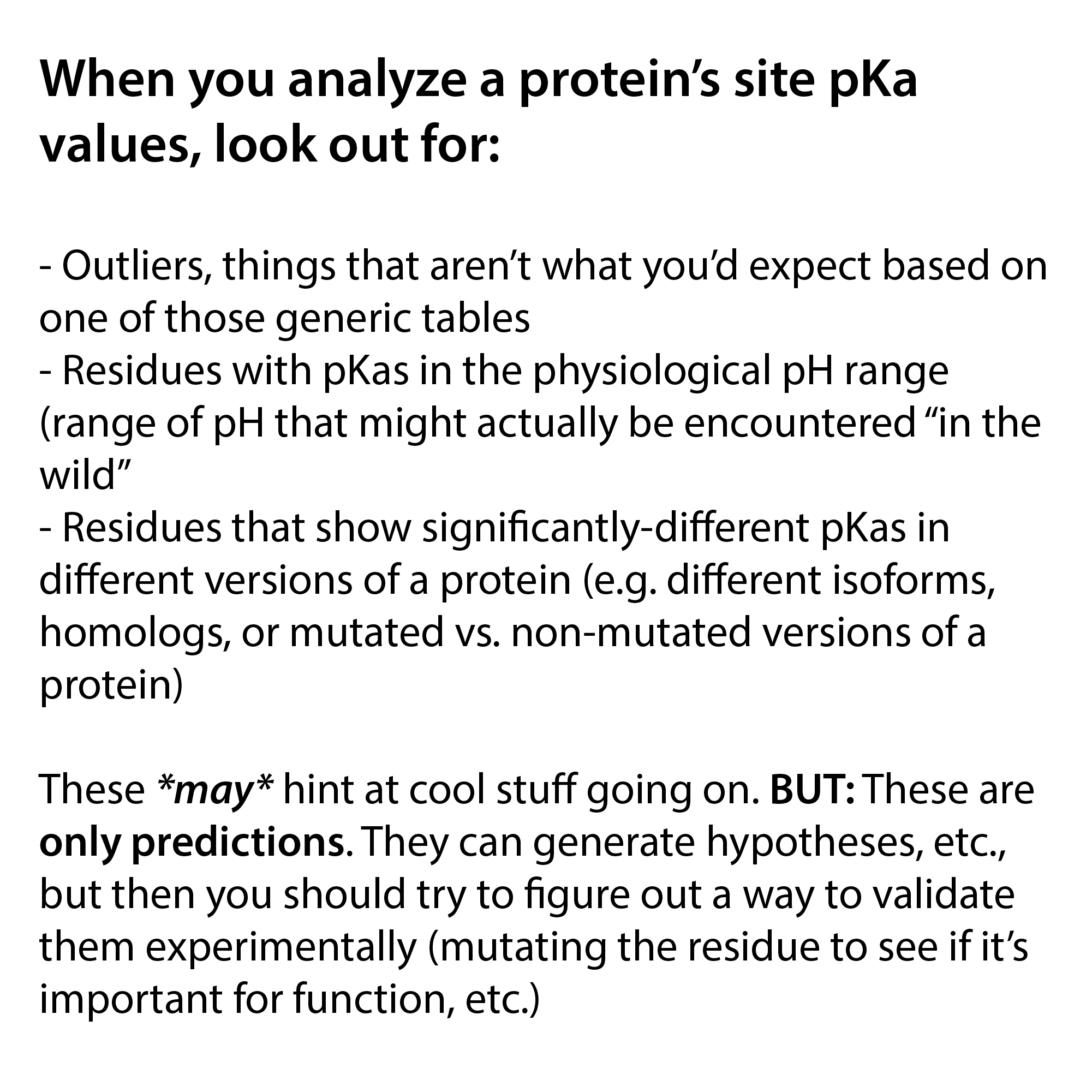
When you analyze a protein’s site pKa values, look out for:
- Outliers, things that aren’t what you’d expect based on one of those generic tables
- Residues with pKas in the physiological pH range (range of pH that might actually be encountered “in the wild”
- Residues that show significantly-different pKas in different versions of a protein (e.g. different isoforms, homologs, or mutated vs. non-mutated versions of a protein)
These *may* hint at cool stuff going on. BUT: These are only predictions. They can generate hypotheses, etc., but then you should try to figure out a way to validate them experimentally (mutating the residue to see if it’s important for function, etc.)
To access H++: http://newbiophysics.cs.vt.edu/H++/index.php
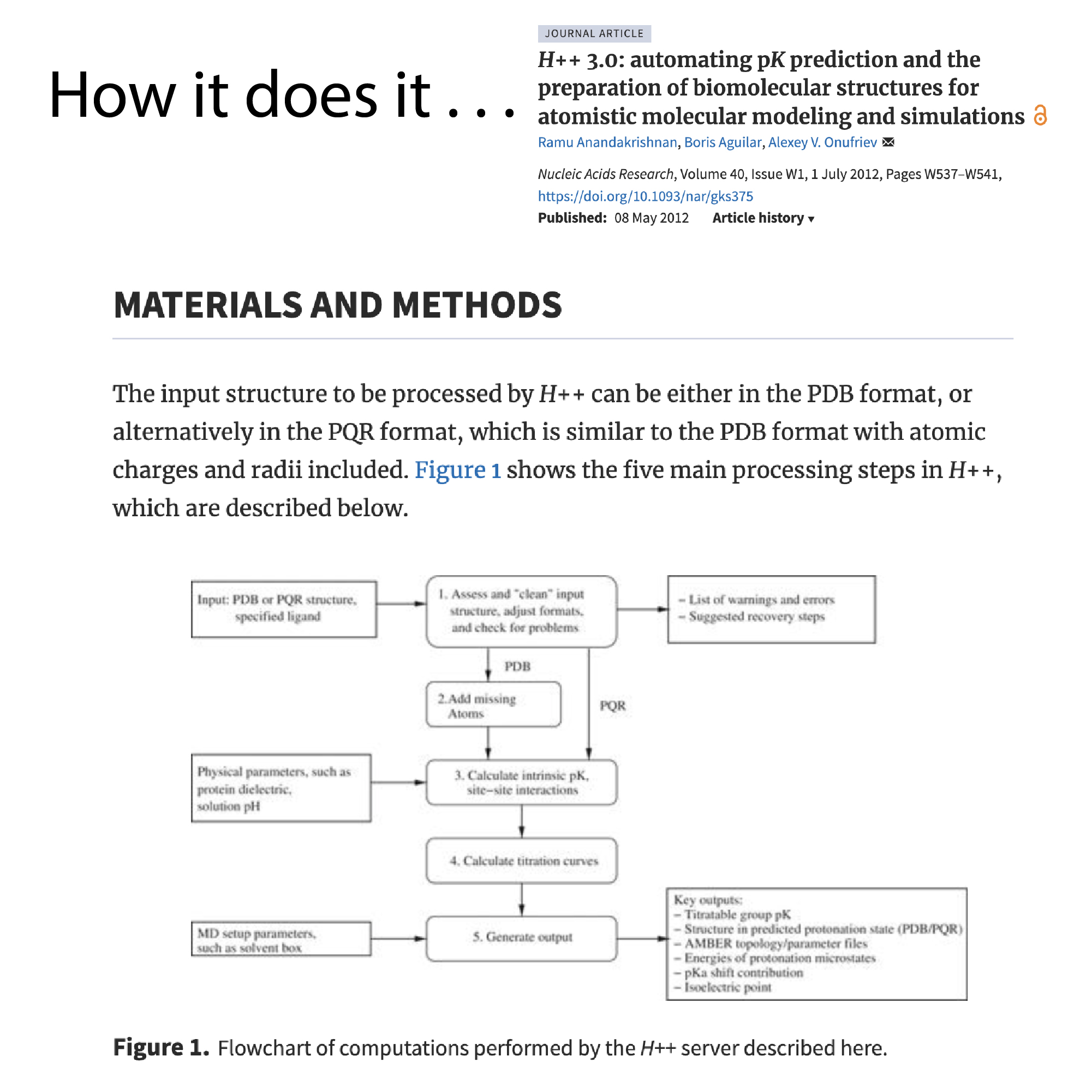
H++ gives you results with a number of different outputs, as I will briefly overview below.
But first, a quick note: You might be able to get a pdb file for your protein from UniProt. If they don’t have one or if you have something custom, you can use AlphaFold to generate one: https://alphafoldserver.com/about
Now, a bit about the results you’ll get. . .
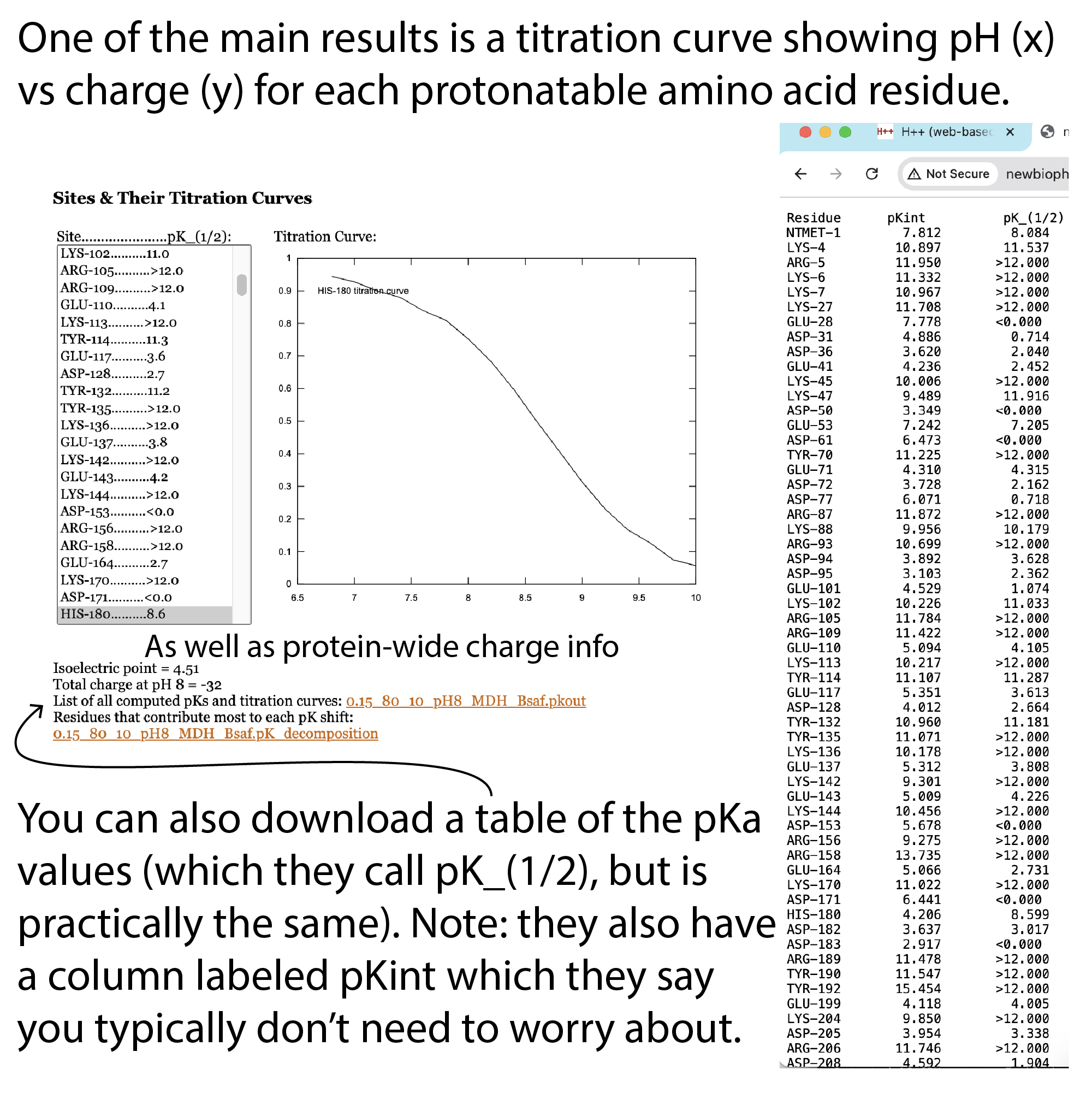
One of the main ones is a titration curve for each protonatable amino acid residue. You can also download a table of the pKa values (which they call pK_(1/2) – see below). Note: they also have a column labeled pKint which they say you typically don’t need to worry about.
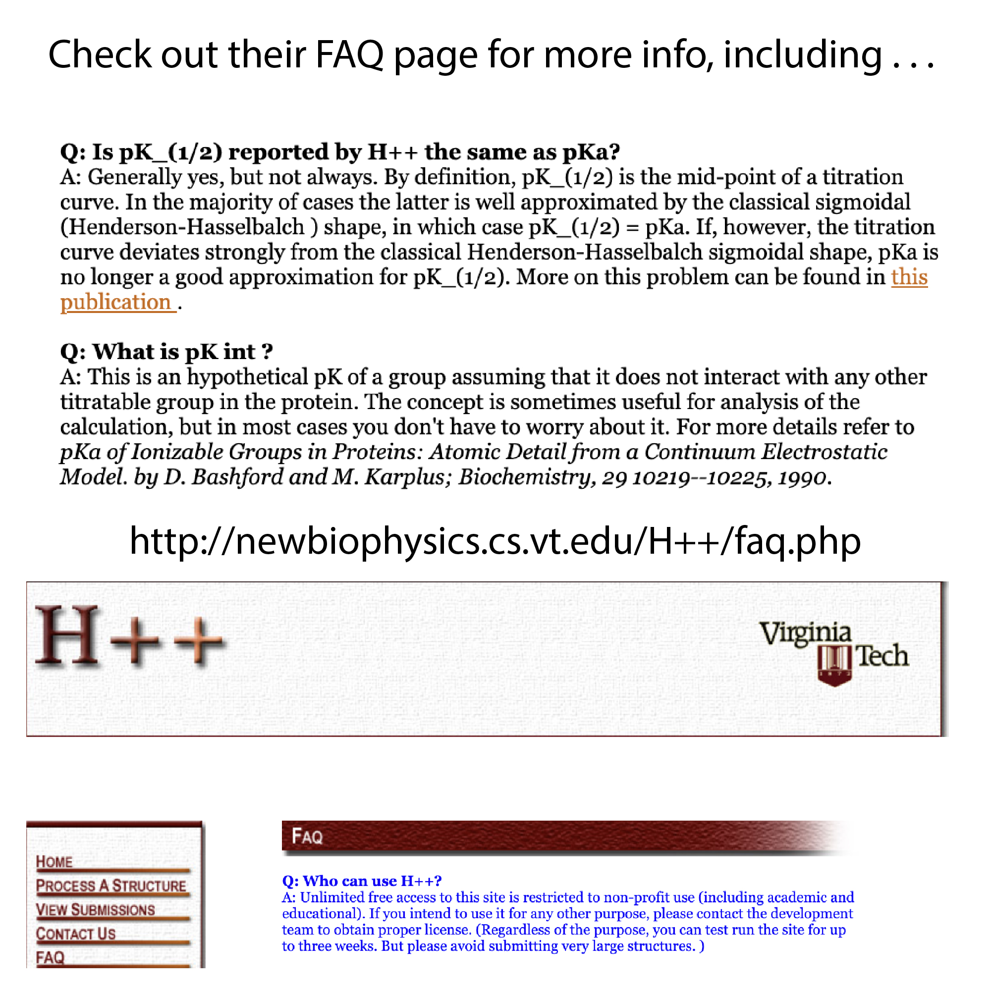
From their FAQ:
“Q: Is pK_(1/2) reported by H++ the same as pKa?
A: Generally yes, but not always. By definition, pK_(1/2) is the mid-point of a titration curve. In the majority of cases the latter is well approximated by the classical sigmoidal (Henderson-Hasselbalch ) shape, in which case pK_(1/2) = pKa. If, however, the titration curve deviates strongly from the classical Henderson-Hasselbalch sigmoidal shape, pKa is no longer a good approximation for pK_(1/2). See Onufriev, A., Case, D. A., & Ullmann, G. M. (2001). A novel view of pH titration in biomolecules. Biochemistry, 40(12), 3413–3419. https://doi.org/10.1021/bi002740q
Q: What is pK int ?
A: This is an hypothetical pK of a group assuming that it does not interact with any other titratable group in the protein. The concept is sometimes useful for analysis of the calculation, but in most cases you don’t have to worry about it. For more details refer to pKa of Ionizable Groups in Proteins: Atomic Detail from a Continuum Electrostatic Model. by D. Bashford and M. Karplus; Biochemistry, 29 10219–10225, 1990. Bashford, D., & Karplus, M. (1990). https://doi.org/10.1021/bi00496a010 “
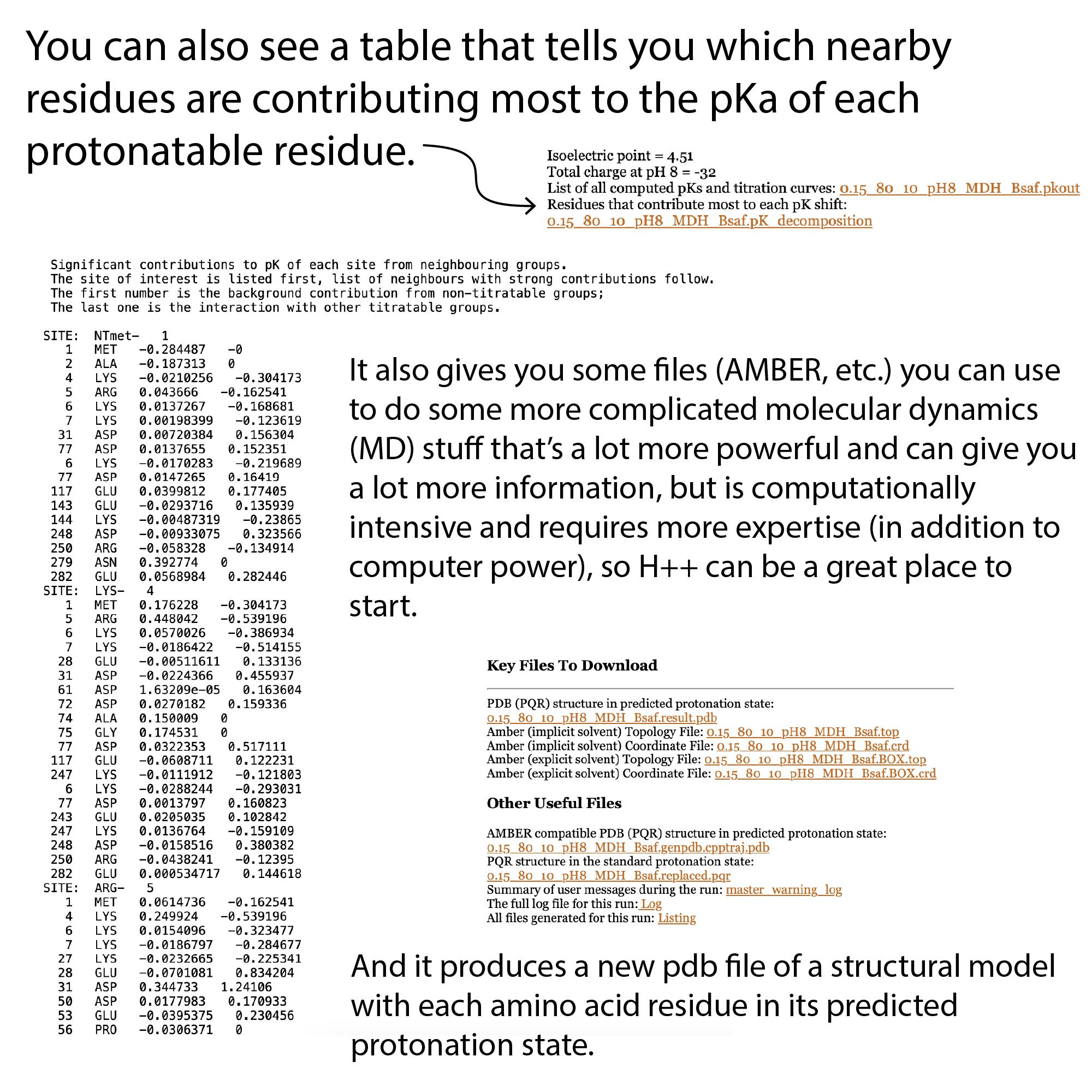
You can also see a table that tells you which nearby residues are contributing most to the pKa of each of those residues.
It also gives you some files (AMBER, etc.) you can use to do some more complicated molecular dynamics (MD) stuff that’s a lot more powerful and can give you a lot more information, but is computationally intensive and requires more expertise (in addition to computer power), so H++ can be a great place to start.
And a new pdb file of a structural model with each amino acid residue in its predicted protonation state.
More in their FAQ: http://newbiophysics.cs.vt.edu/H++/faq.php
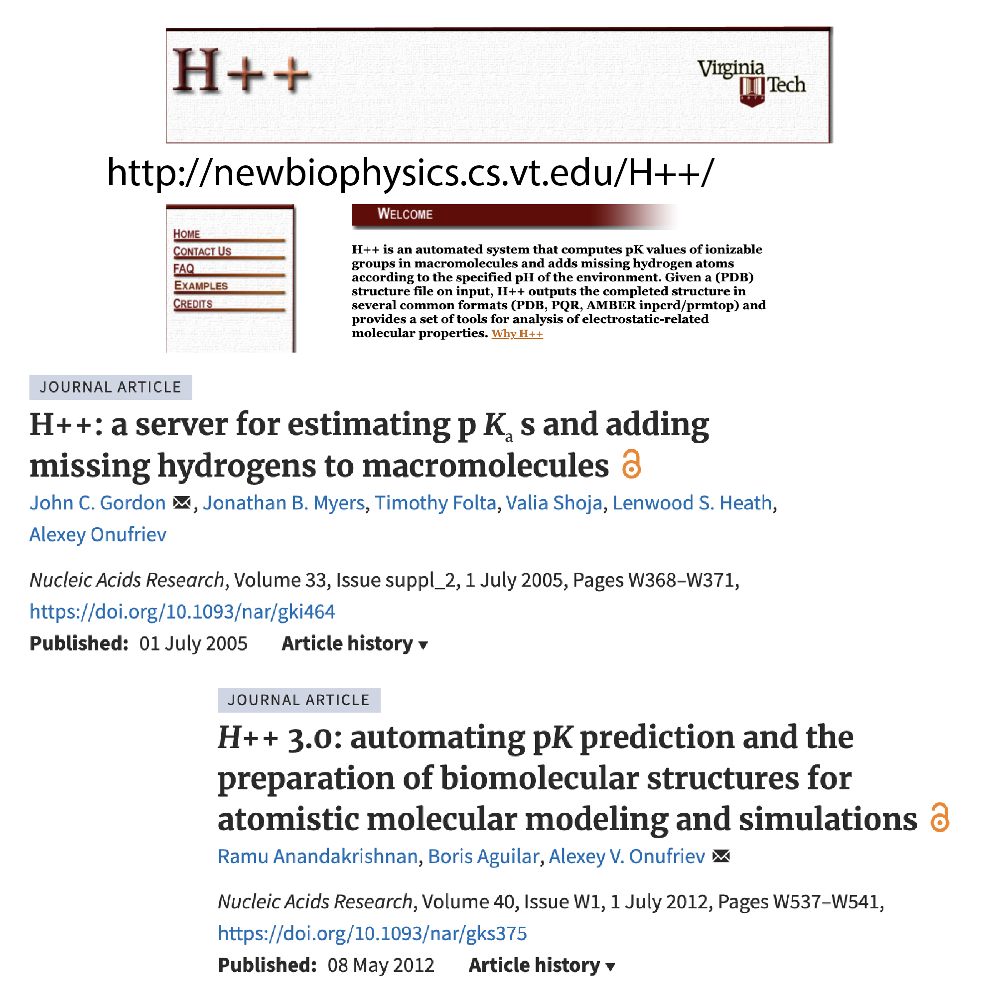
Gordon, J. C., Myers, J. B., Folta, T., Shoja, V., Heath, L. S., & Onufriev, A. (2005). H++: a server for estimating pKas and adding missing hydrogens to macromolecules. Nucleic acids research, 33(Web Server issue), W368–W371. https://doi.org/10.1093/nar/gki464
Anandakrishnan, R., Aguilar, B., & Onufriev, A. V. (2012). H++ 3.0: automating pK prediction and the preparation of biomolecular structures for atomistic molecular modeling and simulations. Nucleic acids research, 40(Web Server issue), W537–W541. https://doi.org/10.1093/nar/gks375
Much more on protein charge: https://bit.ly/isoelectricpoint & https://youtu.be/CLgzYBm_ymk

more background: